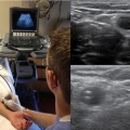
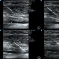
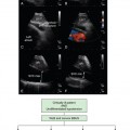
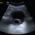
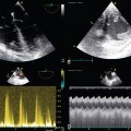
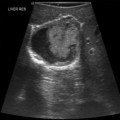
58 Ultrasound is a safe imaging modality and should be considered “first” in many clinical scenarios when imaging is indicated, especially those involving children to avoid the risk for cancer from ionizing radiation (i.e., ultrasound versus computed tomography for the diagnosis of appendicitis).1–3 The cost of ultrasound imaging is also considerably less than that of most other imaging modalities, and it can provide equivalent or better images and thus decrease overall health care cost. When compared with other imaging modalities, ultrasound has the added advantage of providing dynamic examinations, such as evaluation of the shoulder for rotator cuff disease or real-time guidance of procedures such as central line placement and thoracentesis. Ultrasound was introduced into medical student education in the 1990s and proved to be a valuable teaching tool in courses such as anatomy, physiology, and physical examination.4–6 Ultrasound education gradually expanded into the clinical years of medical school, especially in the discipline of emergency medicine.7 The clinical application of ultrasound being taught is as a focused or point-of-care examination designed to answer a specific clinical question such as “Is there a gallstone in the gallbladder that helps explain this patient’s right upper quadrant abdominal pain?” or “Is there overall normal heart function in this patient with shortness of breath?” This point-of-care ultrasound approach has been highly developed in the emergency department setting, and the American College of Emergency Physicians has led the way in defining the necessary training and competencies in point-of-care ultrasound.8 In recent years, ultrasound has been successfully integrated across the entire medical student curriculum. Hoppmann et al reported on 4 years of experience with an integrated ultrasound curriculum.9 As part of the curriculum, all students at the end of the anatomy course in the first year of medical school are observed individually and evaluated for their ability to perform ultrasound on standardized patients and capture and identify anatomic structures in a series of specific ultrasound images. Such structures included the kidney, liver, and Morison pouch in the right upper abdominal quadrant; kidney and spleen in the left upper abdominal quadrant; urinary bladder in the pelvis; carotid artery and internal jugular vein in the neck; and parasternal long-axis view of the heart. The mean percentage of correct responses after 3 years of these examinations was 97.1%. In addition, from student surveys of the same ultrasound curriculum, more than 90% of students thought that the curriculum had enhanced their medical education, allowed increased clinical correlation with basic science instruction, and improved their understanding and skills of the physical examination. Widely used in radiology, cardiology, and obstetrics and gynecology for several decades, ultrasound is now rapidly expanding across many specialties from pediatrics to critical care medicine and orthopedic surgery.10 In addition, the list of clinical applications for ultrasound has grown considerably in recent years and will probably continue to grow as more practitioners explore new applications of ultrasound to improve patient care and safety in their particular specialty.
Ultrasonography: A basic clinical competency
Overview
Ultrasonography in medical student education
Ultrasound across multiple specialties
![]()
Stay updated, free articles. Join our Telegram channel

Full access? Get Clinical Tree


Radiology Key
Fastest Radiology Insight Engine

