14 | Umbilical Cord Complications and Doppler Ultrasound |
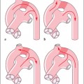
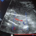
The course and structure of the umbilical cord can, in principle, be displayed by gray-scale ultrasonic imaging when there is sufficient amniotic fluid. However, near term or in critical situations this contrast can be limited, and the abdominal wall may not provide good imaging in all pregnant women. In such cases color-coded Doppler of the umbilical vessels makes it possible to find the umbilical cord, especially when it is in an unusual location, for example, looped around the neck (Figs. 14.1, 14.2a, b), the extremities, or ahead of the presenting part.
The number of vessels can be determined by grayscale or color-coded display.
The thickness of the umbilical cord is of great significance, since it is a measure of the more or less abundant supply of Wharton’s jelly. For one thing, a thick umbilical cord is considered to be an indication of sufficient nutrient supply for the fetus, for another the thickness of the umbilical cord can be an indicator of the degree of risk when there are knots or kinks in the umbilical cord, since a thick cord is hardly in danger of compression.
Color-coded Doppler ultrasound can also detect knots in the umbilical cord, especially when they need to be distinguished from false knots. Doppler ultrasound is also valuable in the detection of eccentric insertions and vasa previa. Similarly, it may be used to make a definitive diagnosis of intertwining of the umbilical cords of monoamniotic twins.
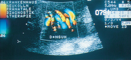
Fig. 14.1 Triple umbilical cord loop. Transverse cut of neck.
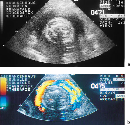
Fig. 14.2 Display of the cord looped around the neck. (a) Horizontal section without color Doppler. (b) Horizontal section with color Doppler.
Doppler Ultrasound Findings when Umbilical Cord Complications Affect Hemodynamics
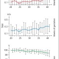
As a rule the unusual findings in the umbilical cord described above do not pose an acute problem at the time of diagnosis. In individual cases, however, spectral or color Doppler sonography may detect a hemodynamic dysfunction by a reduced flow in the umbilical v. or aa. Umbilical cord knots may show a reflection phenomena in the arteries, i. e., the usually smooth waveform of the sonogram of the umbilical aa. shows a notch ig. 14.3). A constriction of the lumen of the umbilical can be seen as a considerable increase in the flow rate and a consequent localized color shift that may extend to aliasing in the color Doppler image.
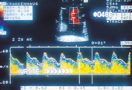
Fig. 14.3 Late systolic notch on the waveform of the umbilical a., possibly caused by a knot in the umbilical cord.
Table 14.1 Elucidation of umbilical cord findings by various ultrasound procedures
Umbilical cord finding | Ultrasound procedures suitable for displaying finding |
|---|---|
Localization | Gray-scale B-mode image/color Doppler ultrasound |
Number of perfused vessels | Color Doppler ultrasound |
Thickness of umbilical cord | Gray-scale B-mode image |
Flow restriction | Doppler sonogram (waveform)/color Doppler ultrasound |
Obstetric Applications of Doppler Ultrasound: References
Diagnostic Doppler Ultrasound in Preeclampsia, Eclampsia, and HELLP Syndrome
Beinder E, Lang N. Veränderungen der mikrozirkulatorischen Reaktivität bei Patientinnen mit Gestose. Gebfra. 1994; 54:268–272.
Brosens IA. Morphological changes in the uteroplacental bed in pregnancy hypertension. Clin Obstet Gynaecol. 1977; 4:573–593.
Campbell S. Nitric oxide: its role in the development of the uteroplacental circulation. 3rd World Congress on Ultrasound in Obstetrics and Gynecology, Las Vegas; 1993.
Campbell S, Diaz-Recasens J, Griffin DR, et al. New Doppler technique for assessing uteroplacental blood flow. Lancet. 1983; 1:675–677.
Campbell S, Pearce JMF, Hackett G, et al. Qualitative assessment of uteroplacental blood flow: early screening test for high risk pregnancies. Obstet Gynecol. 1986; 68:649–653.
Cohen-Overbeek T, Pearce JM, Campbell S. The antenatal assessment of utero-placental and feto-placental blood flow using Doppler ultrasound. Ultrasound Med Miol. 1985; 11:329–339.
Fleischer A, Schulman H, Farmakides G, et al. Uterine artery Doppler velocity in pregnant women with hypertension. Am J Obstet Gynecol. 1986;154:806–813.
Furchgott RF, Zawadski JV. The obligatory role of endothelial cells in the relaxation of arterial smooth muscle by acethylcholine. Nature. 1980; 288:373.
Gonser M, Pfeiffer KH, Dietl J, Hofstaetter C, Gross M. Effect of placental location on uteroplacental Doppler measurements and perinatal risks estimation. J Matern Fetal Invest. 1993; 3:9–13.
Gonser M, Vetter K. Diagnostische und klinische Wertigkeit der Dopplersonographie in der Geburtshilfe. Gebfra. 1995; 55:605–615.
Goodman RP, Killam A, Brash A, Branch A. Prostacyclin production during pregnancy: comparison of production during normal pregnancy and pregnancy complicated by hypertension. Am J Obstet Gynecol. 1982; 142:817.
Hackett G, Cohen-Overbeek T, Campbell S. The clinical value of blood flow studies in obstetric management. In: Jung H, Fendel H, eds. Doppler techniques in obstetrics. Stuttgart: Thieme; pp 58–63.
Harrington KF, Campbell S, Bewley S, Bower S. Doppler velocimetry studies of the uterine artery in the early prediction of pre-eclampsia and intra-uterine growth retardation. Eur J Obstet Gynecol Reprod Bio. 1991; 42 (Suppl.):14–20.
Related posts:
 Diagnosis of the Uterine Tube by Transvaginal Sonography
 Doppler Sonography in Obstetrics—Screening At-Risk Populations
 Doppler Ultrasound Findings Near Term
 Doppler Ultrasound in the Diagnosis of Fetal Anomalies
 Diagnostic Sonography of Blood Flow in Breast Tumors
 Vascular Supply of the Uteroplacentofetal Unit and Techniques for the Examination of Individual Vessels
Stay updated, free articles. Join our Telegram channel

Full access? Get Clinical Tree


