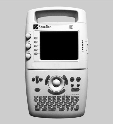
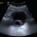
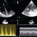
49 During previous conflicts, as recently as the late 20th century, medical diagnostic imaging on the battlefield was mostly limited to sparsely available plain radiography. The introduction of ruggedized handheld ultrasound devices in the late 1990s transformed the diagnostic capability on the modern battlefield.1,2 Although still vulnerable to the harsh environment of war zones, these lightweight portable units now enhance medical care for diverse battlefield settings and medical providers. The use of ultrasound in war zones often reflects its use in traditional hospital environments; however, the unique challenges and constraints of the battlefield give rise to unique applications. Ultrasound imaging for field military use became feasible in 1999 with the release of the SonoSite 180 machine (SonoSite, Bothell, WA), which was developed with a grant from the United States government for military use, with specifications that it be “a field device, hand-held by soldiers or medics, producing high quality images which could be downloaded via satellite to physicians at base hospitals” (Figure 49-1).1,2 To achieve these goals and be practical for field use, designers of this and subsequent ultrasound machines have sought to be lightweight and easy to transport, rugged, and versatile in their power source. War zone ultrasonographers may be equipped with capable technology and basic image acquisition and interpretation training but may lack the expertise to interpret some aspects of the images obtained. Via telesonography, electronic communication tools allow the transmission of ultrasound images from the point of care to specialists for further image interpretation. Several battlefield methods have been described.2–5 Image file capture can be achieved by the ultrasound machine or a secondary device, such as a camera or camera-equipped phone. Image file transmission has been described by phone, landline, and by wireless relay from a portable vest-mounted transmitter to a receiving antenna and then to a satellite.6 The clinical impact of telesonography depends on the speed of image transmission, clarity of the final received image, and the timeliness of pertinent image interpretation. Medical care is delivered to battlefield casualties in several unique settings, from the point of injury to care during evacuation out of the war zone. Each of these settings can pose exceptional challenges to a unique combination of care providers with varying medical skills and familiarity with ultrasound (Table 49-1). These variations allow diverse opportunities for ultrasound to impact the care of injured or ill patients. TABLE 49-1 Characteristics of Different Settings of Care in the War Zone
Use of ultrasound in war zones
Overview
Deployable ultrasound devices and technology
Settings of patient care during war
Setting
Potential Ultrasonographers Providing Care
Challenges to Medical Care
Point of injury
Medics
Soldiers
Enemy fire
Noise
Lack of resources
Lack of medical expertise
Mass casualty events
Prehospital transport
Medics
Enemy fire
Noise
Uncertainty of injuries
Field aid station
Medics
Physicians
Midlevel providers, such as physician assistants
Enemy fire
Uncertainty of injuries
Lack of resources
Lack of medical specialists
Field hospital
Physicians
Midlevel providers, such as physician assistants
Nurses
Medics
Mass casualty events
Variable resources and medical specialists
War zone evacuation
Physicians
Nurses
Medics
Noise
Long transport timesRelated posts:
 Ultrasound-guided peripheral intravenous access
 Ultrasound-guided arterial catheterization
 Endobronchial ultrasound: (CONSULTANT-LEVEL EXAMINATION)
 Ultrasonography in circulatory failure
 Evaluation of right ventricular function in the intensive care unit by echocardiography: (CONSULTANT-LEVEL EXAMINATION)
 Procedural ultrasound for surgeons: (CONSULTANT-LEVEL EXAMINATION)
![]()
Stay updated, free articles. Join our Telegram channel

Full access? Get Clinical Tree


Radiology Key
Fastest Radiology Insight Engine