Figure 24.1.1
Normal premenopausal uterus. Sagittal transvaginal view of the uterus in three women. A: The myometrium (M) is homogeneous and hypoechoic relative to the endometrium (arrowheads). B: The inner myometrium (arrowheads) is less echogenic than the outer myometrium. Both are less echogenic than the endometrium (arrows). C: Arcuate vessels are seen as anechoic serpiginous structures (arrows) in the peripheral aspect of the myometrium.
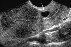
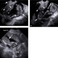
Figure 24.1.2
Nabothian cyst. Sagittal transvaginal view of the uterus reveals a nabothian cyst (arrow) in the cervix.
24.2. Endometrium
Description and Clinical Features
The endometrium is the innermost layer of the uterus that lines the uterine cavity. It has a basal layer adjacent to the myometrium and a functional layer containing glandular tissue. In women of menstrual age, the functional layer changes markedly during the menstrual cycle. The functional layer is shed during menstruation, leaving the thin basal layer as the only remnant of the endometrium. There is often blood and shed tissue in the uterine cavity during this part of the cycle. After menstruation, the endometrium enters its proliferative phase, during which the functional layer regenerates in response to stimulation by estrogen produced by ovarian follicles. This phase continues until ovulation, at mid-cycle, when a dominant ovarian follicle ruptures. The resulting corpus luteum produces progesterone. Under the influence of progesterone, the endometrium enters its secretory phase, during which its glands begin to secrete. The secretory phase continues until menstruation begins anew.
After menopause, the endometrium atrophies. This can lead to vaginal bleeding because the atrophic endometrium is prone to ulceration.
Sonography
In a woman of menstrual age, the sonographic appearance of the endometrium varies during the menstrual cycle corresponding to the anatomic changes described above (Figure 24.2.1). During menses, blood or debris is often detectable in the uterine cavity. In the early proliferative phase, the endometrium appears as a thin echogenic line, corresponding to the apposed thin basal layers. In the late proliferative phase, the functional layer is thicker and hypoechoic, so that the endometrium has a multilayered appearance: a thin echogenic line anteriorly (anterior basal layer), then a thick hypoechoic zone (anterior functional layer), followed by a thin echogenic line in the middle (junction between the two functional layers), then a thick hypoechoic zone (posterior functional layer), and finally a thin echogenic line posteriorly (posterior basal layer). During the secretory phase, the functional layer becomes more echogenic until it becomes isoechoic to the basal layers, so that the endometrium appears as a single thick echogenic band.
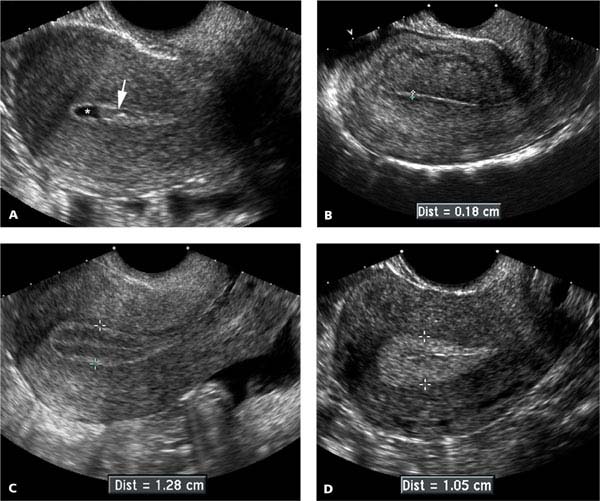
Figure 24.2.1
Normal endometrial appearance during the menstrual cycle. Sagittal midline views of the uterus in women at different stages of the menstrual cycle depict the variations in endometrial appearance. A: Sonogram during menses demonstrates clot (arrow) and fluid (*) within the uterine cavity; the endometrium is seen as a thin echogenic line around the clot and fluid. B: The endometrium (calipers) appears as a thin echogenic line during the early proliferative phase, shortly after menses. C: During the late proliferative phase, the endometrium (calipers) has a multilayered appearance: echogenic around its periphery and in the midline, and hypoechoic in between. D: During the secretory phase, the endometrium (calipers) is thick and echogenic.
Related posts:


Stay updated, free articles. Join our Telegram channel

Full access? Get Clinical Tree


