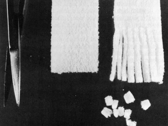
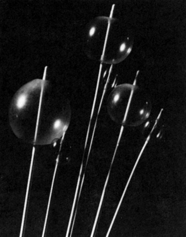
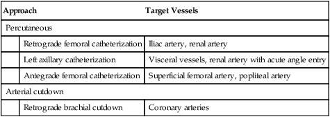
Vascular Interventional Procedures

CHAPTER 17


Vascular Interventional Procedures


The term “interventional radiology” was coined by Wallace in 1976 to describe any selective catheter or needle technique used for the diagnosis and treatment of disease. Although the basic techniques had been used for many years, this subspecialty began in 1964 when Dotter and Judkins successfully recanalized arthrosclerotic stenosed femoral arteries. Drawing on their experience as angiographers and using standard equipment, they were able to pass catheters through occluded arterial segments. Their technique used overlapping (coaxial), or telescoping, catheters to compress the plaque. The method improved, and in 1974 Gruntzig developed the double-lumen balloon sheath catheter, which improved the recanalization procedure and reduced trauma to the vascular system at the puncture site. These and other interventional procedures have proved to reduce the cost of treatment without increasing risk to the patient. They have a low mortality rate and, in many cases, are better tolerated by the patient than are more complicated and extensive therapeutic or surgical procedures. In general, interventional radiography encompasses both vascular and nonvascular procedures. The results of an interventional technique can be diagnostic as well as therapeutic. Samples may also be taken for diagnosis to provide histologic, bacteriologic, and biochemical information.


Current catheter systems used for interventional procedures consist of an arterial guiding sheath catheter. This is a catheter with a thinner wall and larger lumen than the typical angiographic catheter. It acts as a guide for the actual interventional catheter. The sheath also provides the means for the injection of contrast agent as well as performing various hemodynamic measurements.


The catheters used can be of different types: the fixed guide wire and the over the guide wire catheter system. Each of these systems has advantages and disadvantages. The fixed guide wire system has the balloon directly attached to a guide wire equipped with a flexible tip. This type of system has the advantage of being able to be manipulated by a single person. Its disadvantage is that it must be completely removed if another size or type of catheter is to be inserted. In the over the guide wire catheter system the catheter rides over a preplaced guide wire. It is a bit more cumbersome to use than the previous system but has increased flexibility in the types of catheters and guide wires that can be accommodated. In this type of system the guide wire remains in its location, facilitating the removal of one catheter and replacement with another without retracing the route with a new guide wire.


There are four major divisions of vascular interventional procedures: those used to reduce or stop blood flow, procedures used to increase blood flow by dilating or recanalizing occluded vessels, interventions used to remove foreign bodies from the vascular system, and other procedures. The first three categories of interventions can be applied in the vasculature of the major organ systems of the body. Certain procedures such as TIPS (transjugular intrahepatic portosystemic shunting) are used in specific organ systems and fall in the fourth category, “other interventions.” The means of vessel access for these procedures is usually the percutaneous method originally described by Seldinger. Depending upon the target, access may be either arterial or venous.


Box 17-1 illustrates the present scope of interventional radiography for both vascular and nonvascular procedures. The nonvascular interventional procedures will be discussed in Chapter 19 but are listed here for comparison with the vascular interventions. Vascular interventions can be divided into methods that either reduce or increase blood flow. These techniques are meant to be therapeutic in nature. Vascular interventional radiography procedures use angiography as the primary procedural method for access to the site of the pathologic process. In many cases the percutaneous interventions can save the patient from undergoing a lengthy surgical procedure.


TECHNIQUES USED TO REDUCE BLOOD FLOW


Procedures for reducing blood flow includ embolization and balloon occlusion, intravascular infusion of vasoconstrictors, and intravascular electrocoagulation. Most of these techniques are performed using percutaneous puncture of the femoral or other superficial arteries. Occasionally, the embolic material is inserted directly into the vessel via an arteriotomy.


Indications and Contraindications


Transcatheter embolization is indicated in cases of posttraumatic hemorrhage, for occlusion of the blood supply to highly vascular neoplasms, and for reduction of bleeding during and after surgical procedures. Because transcatheter embolization is presently used in high-risk cases, there are nodefinitive contraindications to the procedure. Ideally, the embolization method chosen reduces the flow of blood without incurring ischemia of the tissue.


The occlusion of blood vessels can also be accomplished using an electric current to stimulate thrombus formation. Electrocoagulation can be used in cases of diagnosed tumors, hemangioma, and arteriovenous fistulas.


Infusions of vasoconstrictors are indicated in cases of suspected upper gastrointestinal bleeding and as an aid to improving the diagnostic capabilities of angiography in certain areas of the body. If vasoconstrictors are administered during hepatic angiography or peripheral angiography, the diagnostic value of the examination is enhanced. In cases of suspected hepatoma and adenoma, the instillation of vasoconstrictors results in an improved visualization of the neoplasms.


Procedure


Transcatheter Embolization and Balloon Occlusion


Transcatheter intravascular occlusion, the process of closing off or obstructing the inside of blood vessels, is accomplished by placing a foreign substance, tissue, or blood clot into the lumen of a selected vessel through a catheter. Naturally occurring particulate types of embolic materials such as autogenous and autologous blood clots and tissue fragments (muscle, fat, and so on) have become obsolete with the increased use of synthetic agents that can be delivered through percutaneous catheter methods. The following is a list of the embolic materials that are used for vascular embolization:


The search for the ideal embolic material has yielded many different products, but none exhibits all of the characteristics of an ideal substance. An ideal embolic material is nontoxic, stable, insoluble in the vascular system, radiopaque, and capable of being shaped, sterilized, and introduced via an angiographic catheter. The material used is determined by the pathology, the patient’s clotting ability, and the nature of the blood supply. Occasionally, it is necessary to use a combination of more than one type of embolic material to successfully isolate an area.


The substances used to occlude the blood vessels can be categorized by how long they remain in the vessel—they can be temporary or long lasting. Substances such as Gelfoam, autologous blood clots, muscle tissue clots, and balloon catheters are temporary occluders. These substances are either mechanically removable, such as the balloon catheter, or quickly absorbed by the body through intravascular lysis.


Embolic media such as Gianturco coils, detachable balloon catheters, isobutylcyanoacrylate (IBCA), and polyvinyl alcohol foam are long-term agents used for permanent occlusion of vessels.


Cellulose Sponge Embolic Media.

These media include microfibrillar collagen,oxidized cellulose, and Gelfoam, which is the most popular. Gelfoam is manufactured in sheets and in powder (Fig. 17-1). It was first used in 1945 to control hemorrhage during neurosurgery, and it has been successfully used in intravascular occlusion. The powdered form consists of particles ranging from 40 to 60 μm in size. When Gelfoam is used, small fragments (pieces) are cut from the sheet and soaked in normal saline solution, a contrast agent, or both to provide radiopacity. These pieces are then placed into the vessel by injection through an angiographic catheter. The size of the inside lumen of the catheter determines the maximum size of the piece to be used. Gelfoam has the disadvantage of being easily refluxed, causing recanalization of the vessel. This type of embolic medium, when injected into the lumen of a vessel, provides a framework for blood clot formation. Intravascular lysing of this type of material can occur in a relatively short period, but it is much more persistent in a vessel than an autologous blood or tissue clot alone.


Gelfoam powder that has been radiopacified by the addition of tantalum powder is used to produce occlusion at the precapillary level. At this level, recanalization by collateral vessel formation is minimized, making Gelfoam powder an excellent choice for tumor treatment.


This type of embolic medium lasts from less than 48 hours to 30 days.


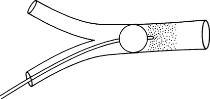
Balloon Catheter Occlusion.

Balloon catheters provide a safe, temporary method of occluding vessels to control or prevent bleeding, as well as isolating vessels for selective infusion of chemotherapeutic agents or placing other types of embolic media (Fig. 17-2). The advantages of using the balloon catheter are that it is retrievable for removal and the position of the balloon can be changed easily.


Four types of balloon catheters are available. The single-lumen type is used with catheter introduction and exchange sheath sets or coaxial catheters. The double-lumen type is constructed to permit contrast injection and balloon inflation in the same catheter. The double-lumen catheter consists of one catheter within a second, larger catheter. The third type—flow-directed detachable miniature balloon catheters—are used for permanent occlusion of high-flow vascular lesions. The controlled-leak balloon catheter systems are also flow-directed catheters that allow the delivery of a liquid embolic material or contrast agent.


Single- and double-lumen catheters are primarily used as temporary occluders. Detachable balloon catheters are used to provide long-term occlusion, especially in areas in which high blood flow is present. Detachable balloon catheters are equipped with self-sealing valves and are usually filled with contrast medium or another opaque medium when the study is performed. The catheter has a stainless-steel stem that fits into the valve of the balloon. When the balloon is placed in the vessel to be occluded, it can be inflated. The catheter is then withdrawn, disconnecting the stem and leaving the balloon in the vessel. Detachable balloons can also be flow-guided into position. The detachable balloon catheters are used with coaxial catheter systems to facilitate placement of the miniballoon.


A balloon catheter can be used in conjunction with embolic material and in such cases function like a cork to prevent reflux of the particulate material from the target vessel. Once the particulate embolic material has been firmly set in place, the balloon can be removed (Fig. 17-3).


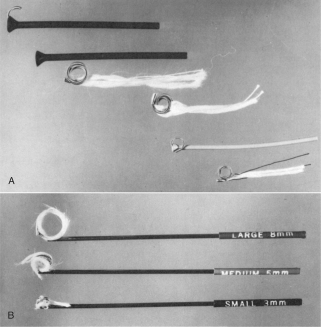
Occluding Spring Emboli and Other Mechanical Occluders.

Occluding springs are mechanical devices of stainless-steel coiled wire about 5 cm long and available in 3-, 5-, 8-, 10-, 12-, and 15-mm diameters. Strands of wool, silk, or Dacron are affixed to the wire to provide a framework for clot formation. The wool-tailed coils are primarily used for larger arteries, whereas the silk- or Dacron-tailed coils are used to occlude smaller arteries. These coils were developed in 1975 by Gianturco, Anderson, and Wallace and are commonly called Gianturco-Anderson-Wallace (GAW) coils (Fig. 17-4).


These devices are supplied uncoiled and braided into an introducer cartridge. To place the coil into the catheter, the introducer cartridge is inserted into the stopcock until it contacts the flared catheter end. The coil is then pushed into the catheter with the stiff end of a guide wire. The distance that the coil should be pushed into the catheter in the loading stage is usually recommended by the manufacturer. In the case of the GAW coil, the manufacturer (Cook, Inc.) recommends leading for a distance of 20 to 30 cm. The guide wire and introducer are removed, and the spring embolus is then pushed through the distal end of the catheter for placement in the vessel. A smaller version of the GAW coil, the minicoil, is also available. This unit is delivered through a 5F catheter.


Occluding spring emboli are considered permanent embolic media because they remain in the vessel for more than 30 days.


The bristle brush has also been successfully used to occlude vessels. This device resembles a miniature chimney cleaning brush and has a central core consisting of a stainless-steel coil with nylon pieces threaded through it. These brushes require the use of large-bore catheters for introduction into the target vessel, where they remain to provide a framework for clot formation. They also are considered permanent embolic media.


Liquid Embolic Media.

Substances in this category include tissue adhesives (polymerizing agents) such as IBCA and silicon elastomer. These products are long-term embolic media, lasting more than 30 days. These materials are used primarily in life-threatening cases. They solidify (polymerize) when they come into contact with ionizing solutions. Unlike other embolic materials, these substances do not form the framework on which a blood clot is produced; rather, they form a permanent embolus that seals the vessel almost immediately.


IBCA is used for direct vessel occlusion but has not yet been approved by the Food and Drug Administration (FDA) for unrestricted use. A permit must be secured for any use of this material in human beings.


The silicon elastomer embolic medium is a low-viscosity substance that exhibits a more controlled polymerization time than IBCA, making it a better choice for occlusion of extensive arteriovenous malformations. Silicon elastomer can be mixed with iron microspheres, which allows an externally applied magnet to control the position of the material until the polymerization process is complete. This procedure was devised to prevent the embolic material from moving to a nontarget vessel.


The technique for placement of these liquid emboli substances requires the use of coaxial catheters. These are specially paired catheters comprising a larger outer catheter and a smaller inner catheter. These sets come with a correctly sized guide wire and are complete for use during intravascular embolization procedures (Fig. 17-5). The large outer catheter is passed through the vessel to the approximate site of occlusion. A small amount of sterile silicon grease can be applied to the tip of the small inner catheter, which is then passed through the outer catheter into the specific vessel to be occluded. The application of the silicon grease helps to prevent adhesion of the catheter tip to the vessel wall after injection of the embolic medium.


The liquid tissue adhesive, mixed with a contrast agent, is then injected into the site. It is important for the inner catheter to be removed as soon as the injection is complete to avoid adhesion of the catheter tip to the vessel wall. Because the substances polymerize when in contact with an ionic solution, the catheters cannot be flushed with normal saline solution. The flushing may be accomplished with a 10% solution of dextrose and water.


Embolization by percutaneous catheter delivery of absolute ethanol has proved to be safe and efficient. Its main mode of action is by damaging the endothelium and stimulating the coagulation systems that cause the vascular occlusion. It is generally used with one of the nondetachable balloon systems. The balloon helps to control reflux of the absolute ethanol. This substance has several advantages such as availability, lack of toxicity, low cost, and low complication rate. Ethanol can also be mixed with one of the nonionic contrast agents to monitor and control the embolization procedure.


Routine angiography is usually performed at the beginning of the procedure to localize the site of bleeding and again after injection of the material to confirm occlusion of the vessel. Use of liquid embolic substances can cause a mild histiocytic reaction in the vessel lumen that does not involve the vessel walls or adjoining parenchyma.


Intravascular Electrocoagulation


Intravascular electrocoagulation, as the name implies, produces a thrombus in the vessel as a result of the application of a direct electric current. Its use was initially limited because of the problems associated with the technique. Early electrocoagulation devices used a stainless steel anode that was subject to destruction due to the passage of an electric current (electrolysis). The breakdown of the stainless steel often caused the anode tip to become detached inside the vessel. The use of stainless steel and the length of time required to produce a clot in larger vessels restricted the procedure to the occlusion of vessels less than 5 mm in diameter. The lengthy procedure resulted in tissue injury from the heat of the ground plate that was placed in contact with the patient. Modern devices use a platinum anode that resists the corrosive effects of electrolysis and a special lubricated grounding pad that prevents skin burns. This allows thrombus formation in larger vessels while protecting the patient from mechanical injury.


The Guglielmi detachable embolization coil is an example of a device that combines electrocoagulation with a detachable mechanical device to achieve satisfactory results.1 Once in place, the electric current promotes thrombus formation, and detachment of the coil aids in the occlusion process through electrolysis of its soft stainless-steel connector. The procedure time is shortened, and the device can be manufactured in various lengths and coil sizes for use in a variety of vessel lumens.


Transcatheter Infusion Therapy


The slow infusion of vasoconstrictors such as vasopressin into a vessel through a catheter is a useful therapeutic technique for the control of bleeding. Vasoconstrictors cause contraction of the muscles associated with the arteries and capillaries, resulting in an increase in the resistance to the flow of blood with a corresponding increase in blood pressure.


Transcatheter infusion therapy is used extensively in the gastrointestinal tract to create controlled ischemia. Transcatheter infusion of vasopressin is made selectively at a rate of 0.2 unit per minute for 20 minutes followed by a repeat angiogram. The dose can be increased to 0.4 unit per minute for an additional 20 minutes if bleeding continues; however, if the bleeding does not stop at this dose infusion, therapy should be discontinued and alternative embolization procedures used. If the bleeding is successfully controlled, the dose should be continued for 12 to 24 hours followed by a 50% reduction in dose for an additional 24 hours. The patient should remain stable for at least 12 hours after the treatment before the catheter is removed. During this period, an infusion of normal saline or dextrose solution should be administered.


In most cases, the treatment will stop arterial or mucosal bleeding without any further intervention. Selective infusion of vasoconstrictors is also a valuable adjunct to mechanical transcatheter embolization by helping to prevent the reflux of the embolic material during placement of the embolic material.


The most popular pharmacologic agent currently used for this procedure is vasopressin. It is supplied as an aqueous solution of the substance pressor that is synthesized from the posterior pituitary gland. Vasopressin has been very successful when used to control bleeding in the gastrointestinal tract, primarily because it causes bowel constriction as well as arterial constriction in this anatomic area. Both of these actions combine to provide a reduction in the flow of blood, allowing clot formation at the site of the bleeding. Other agents such as norepinephrine and propranolol have also been investigated, but the complication and reaction rates are higher than those with vasopressin. These agents also do not create a sustained effect and have not been extensively used.


Complications


Use of transcatheter vascular embolization produces certain reactions, including localized transient pain accompanied by a mild fever, malaise, increased pulse and respiration rates, restlessness, irritability, loss of appetite, and insomnia for a period of 24 to 48 hours after embolization. Other complications may occur, such as embolization of nontarget organs, because of reflux of the embolic medium, ischemia, nerve palsy, infection, and possibly death. It should be remembered that these procedures are performed on patients who present a great surgical risk and on patients who are in life-threatening situations. As with any radiographic procedure, it should be determined that the benefit of performing any interventional procedure outweighs the risks presented by the procedure.


Intravascular electrocoagulation can cause damage to the vessel in which the thrombus is formed. Perforation is another risk associated with this technique.


When a vasoconstrictor such as vasopressin is used to control bleeding, it is important to monitor the patient because vasopressin is a potent diuretic that can cause water retention or have a direct depressive effect on the myocardium.


TECHNIQUES USED TO INCREASE BLOOD FLOW
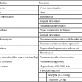

Interventional procedures can also be applied to increase the flow of blood in a particular vessel. Four types of procedures are used: percutaneous transluminal angioplasty (PTA), vascular stent placement, intravascular thrombolysis, and intra-arterial infusion of vasodilators. These procedures provide relatively safe methods of increasing blood flow without the necessity of extensive surgery. These studies are usually done using the percutaneous approach. The arterial cut-down approach is occasionally used, and the necessary sterile tray should be available in the radiography suite. Box 17-2 summarizes some common approaches used for angioplasty.


Indications and Contraindications


Interventional procedures for increasing blood flow are used to either dilate stenotic vessels or recanalize obstructed vessels. Percutaneous transluminal angioplasty (PTA) encompasses both dilation and recanalization. It is used to treat stenotic arterial disease primarily in medium- to large-sized vessels. Coronary, renal, and peripheral arteries are prime candidates for PTA. The lesions treated are usually localized and contain either fibromuscular or complicated plaque (atheroma). Fibromuscular plaque affects the intima by the production of smooth muscle cells, collagen, elastic fibrils, and some lipids. Complicated plaque is actually a fibromuscular plaque encased in a thin fibrous cap. The lesions should be short (not more than 10 cm) for successful angioplasty. Congenital coarctation, Takayasu’s arteritis, and fibromuscular dysplasia can also be treated with PTA.


One drawback to PTA is the potential for restenosis of the vessel by the procedure due to intimal hyperplasia or elastic recoil of the vessel wall. The use of intravascular stents has reduced the negative aspects of percutaneous transluminal balloon angioplasty and provides a method of maintaining the dimension of the vessel lumen as well as its overall patency.


Intravascular thrombolysis is used to treat arterial thromboemboli. The areas that benefit most from this technique are the coronary, peripheral, visceral, and pulmonary vascular sites.


Infusion of vasodilators is useful in treating cases of vessel spasm or constriction and has been successful in the treatment of atherosclerosis obliterans, a common cause of vascular occlusion.


There are no defined contraindications to the use of these procedures. In most cases, the patients are usually candidates for surgery, and attempts at treatment with these techniques usually outweigh the risks involved. When percutaneous transluminal coronary angioplasty is done, a surgical team and an operating room should be available for immediate coronary bypass surgery if complications occur. The only other contraindication is if the anatomy precludes the passage of a dilation catheter.


Procedure


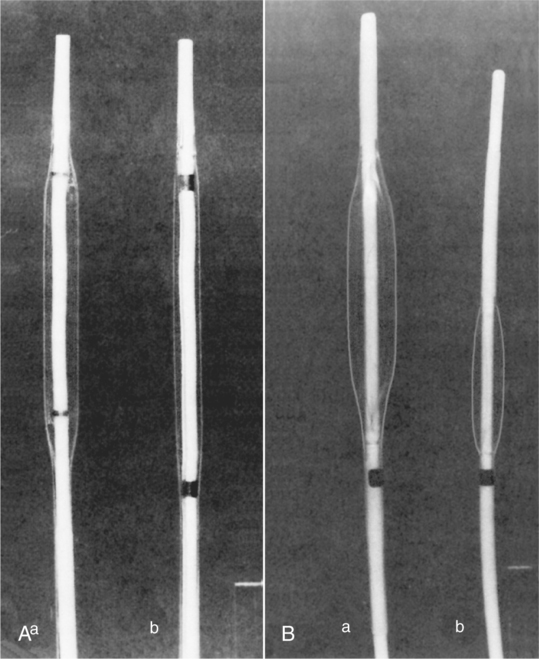
Percutaneous Transluminal Angioplasty


Percutaneous transluminal angioplasty is a nonsurgical procedure defined as the process of accessing a blood vessel through a puncture in the skin and performing a procedure through the blood vessel to reshape or repair the inside diameter of the vessel through balloon inflation. This procedure was first performed in 1964 by Dotter and Judkins; they used a coaxial catheter system (telescoping catheter) to recanalize a stenosed femoral artery. In 1974, Gruntzig developed the double-lumen balloon catheter, which reduced the complication rate at the puncture site. Currently, there are many different balloon lengths and diameters available for use during PTA (Fig. 17-6).


Stay updated, free articles. Join our Telegram channel

Feb 27, 2016 | Posted by in GENERAL RADIOLOGY | Comments Off on Vascular Interventional Procedures

Full access? Get Clinical Tree

Get Clinical Tree app for offline access