and Vincent L. Sorrell2
(1)
Division of Nuclear Medicine and Molecular Imaging Department of Radiology, University of Kentucky, Lexington, Kentucky, USA
(2)
Division of Cardiovascular Medicine Department of Internal Medicine Gill Heart Institute, University of Kentucky, Lexington, Kentucky, USA
Electronic supplementary material
The online version of this chapter (doi: 10.1007/978-3-319-25436-4_14) contains supplementary material, which is available to authorized users.
There are several etiologies for abnormal findings related to the peripheral or central vascular system in the chest (Table 14.1). Extravasation may occur during intravenous injection of the radiopharmaceutical. Depending on the amount extravasated, the radiopharmaceutical may be picked up by the lymphatics and deposited in the regional lymph node basin, typically the axilla after an injection in the upper extremity (Fig. 14.1). When extravasation is suspected, it might be prudent to image the injection site to ensure complete intravascular delivery of the radiopharmaceutical. An incomplete injection, particularly during the stress portion of the examination, may result in spuriously normal-appearing (false-negative) myocardial perfusion due to an inadequate ratio of administered activity relative to the earlier rest injection (Burrell and MacDonald 2006; Chamarthy and Travin 2010; Gedik et al. 2007; Gentili et al. 1994; Howarth et al. 1996; Strauss et al. 2008). Extreme extravasations can cause scatter artifact, reiterating the importance of raising the patient’s arms out of the field-of-view. Additionally, other maneuvers such as shielding can be employed when appropriate.


Table 14.1
Differential diagnosis of “hot” and “cold” imaging findings related to the vascular system
Organ system | “Hot” finding | “Cold” finding | References |
|---|---|---|---|
Vascular system | Contamination during injection Extravasation at injection site Retention in central veins Retention in central venous catheters/ports Pulmonary arterial wall | Dilated pulmonary arteries | Aras et al. (2003) Burrell and MacDonald (2006) Chamarthy and Travin (2010) Gedik et al. (2007) Gentili et al. (1994) Howarth et al. (1996) Shih et al. (2002) Strauss et al. (2008) Tallaj and Iskandrian (2000) |


Fig. 14.1


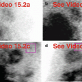
Marked radiopharmaceutical extravasation at right arm injection site with visualization of right axillary lymph node. This occurred during the rest portion of the one-day rest/stress MPI protocol (a–c). The patient’s right arm was positioned alongside the right chest wall for imaging. Note the significant “streaky” scatter effect onto the patient’s right chest wall and abdomen (a). The injection site can be more clearly defined by adjusting the contrast settings during review (b, c). The extravasation is still evident on the later stress imaging (d, e, f). Although it can be appreciated on the rest raw data (a), the right axillary lymph node is more clearly defined with time (e). The scatter effect is still present, although somewhat mitigated (f). However disconcerting on the raw data (a, d), there is no significant detrimental effect on the processed SPECT images (g) nor the gated SPECT images (h), and the examination is reportable. There is a small, mild, fixed inferior-lateral-basal defect, probably artifactual. If the extravasation had occurred during the stress test, the MPI would have been compromised because the desired 3:1 ratio of stress-to-rest administered activity would not have been achieved. A 50-year-old male is on dialysis for diabetes-related end-stage renal disease, has hypertension, and had coronary artery bypass surgery (CABG).(a) Rest raw projection images at usual contrast setting to view heart (Video 14.1a, frame 1), 99mTc sestamibi. (b) Rest raw projection image (Video 14.1b, frame 20) adjusted to show relative intensity of injection site to heart, 99mTc sestamibi, injection site (blue box), heart (pink box), gallbladder as reference point (yellow box). (c) Rest raw projection image (Video 14.1b, frame 20) adjusted to show focality of very “hot” injection site (blue box) in right arm without blooming artifact, 99mTc sestamibi. (d) Stress raw projection images at usual contrast setting to view heart (Video 14.1c, frame 1), 99mTc sestamibi. (e) Stress raw projection images at usual contrast setting to view heart (Video 14.1d, frame 9), 99mTc sestamibi, right axillary uptake (blue box). (f) Stress raw projection image (Video 14.1d, frame 16) at usual contrast setting to view heart, 99mTc sestamibi, injection site (blue box), scatter artifact (red box). (g) Stress/rest processed SPECT images (SA, HLA, VLA). (h) Stress gated SPECT images (Video 14.1e, frame 1) (SA, VLA, HLA)
Stay updated, free articles. Join our Telegram channel

Full access? Get Clinical Tree


