Small vessel: Henoch-Schönlein purpura, lupus vasculitis, Behçet disease, Wegener granulomatosis
IMAGING
• Takayasu arteritis
Classically involves aortic arch
Wall thickening of vascular segment in acute phase
– Can mimic atherosclerotic disease (particularly in mesenteric vessels), but typically smooth, regular, and encompasses longer segment
Chronic stenoses with post-stenotic dilatation, aneurysms, occlusions, and collateral vessel formation
• Polyarteritis nodosa
Involves bifurcations of medium and small sized arteries with branch-point aneurysms
Renal and mesenteric vessels most often involved
Renal infarction and atrophy with striated nephrograms
• Henoch-Schönlein purpura
GI tract often shows manifestations of ischemia
Bowel wall thickening, narrowing, and intussusceptions
Intussusceptions very common in pediatric population
Extensive abdominal inflammation
• Wegener granulomatosis
Kidneys are involved in 80% of cases
Microaneurysms with renal parenchymal scarring, hemorrhage, and bowel ischemia
• Lupus vasculitis
At risk for bowel complications/ischemia due to vasculitis and hypercoagulability (antiphospholipid syndrome)
• Behçet disease
Most often involves distal ileum and closely mimics Crohn disease or malignancy
DIAGNOSTIC CHECKLIST
• CT findings that resemble bowel or renal ischemia in a young person should raise concern for vasculitis
(Left) Axial CECT in a 21-year-old woman with severe abdominal pain shows wedge-shaped defects in the kidneys representing acute ischemic injury.
(Right) Axial CECT in the same patient shows long-segment bowel wall thickening and submucosal edema , findings compatible with enteric ischemia. Rheumatoid vasculitis was subsequently confirmed. Findings that suggest bowel or renal ischemia in a young patient, as in this case, should raise suspicion for vasculitis.
(Left) Sagittal CECT demonstrates diffuse narrowing of the abdominal aorta below the superior mesenteric artery with surrounding soft tissue thickening .
(Right) Coronal CECT in the same patient nicely demonstrates the narrowing and thickening of the abdominal aorta extending to involve the common iliac arteries. This is a common appearance for a large vessel vasculitis (giant cell vasculitis) with active inflammation.
TERMINOLOGY
Definitions
• General term describing a diverse group of diseases characterized by inflammation/necrosis of blood vessels
Classified by size of blood vessel involved into small vessel, medium vessel, and large vessel vasculitis
Large vessel: Takayasu arteritis
Medium vessel: Polyarteritis nodosa
Small vessel: Henoch-Schönlein purpura, lupus vasculitis, Behçet disease, Wegener granulomatosis
• Takayasu arteritis: Chronic granulomatous inflammatory vasculitis affecting aorta and its main branches
• Polyarteritis nodosa: Fibrinoid necrotizing vasculitis involving small and medium vessels with formation of multiple branch-point aneurysms
• Henoch-Schönlein purpura: Hypersensitivity-related acute vasculitis affecting small vessels with deposition of IgA-complexes in skin, joints, kidneys, and GI tract
• Wegener granulomatosis: Granulomatous vasculitis of respiratory tract and kidneys
• Lupus vasculitis: Complex autoimmune disease with associated necrotizing vasculitis affecting small vessels
• Behçet disease: Necrotizing vasculitis of small vessels affecting multiple organs
IMAGING
Imaging Recommendations
• Best imaging tool
CT angiography: First-line modality (regardless of size of vessel involved)
– Allows visualization of vascular abnormalities and extravascular organ involvement
Conventional angiography helpful in equivocal cases
Duplex ultrasound: Screening test for stenosis of proximal mesenteric/renal arteries based on velocity criteria
PET/CT: FDG-avidity of involved vessels and visceral sites
General Features
• Best diagnostic clue
Takayasu arteritis
– Irregularity, stenosis, or inflammatory wall-thickening of aorta or main aortic branches on angiography or CT
Polyarteritis nodosa
– Multiple aneurysms in renal and mesenteric arteries
Henoch-Schönlein purpura
– Multifocal bowel wall thickening and luminal narrowing on CT in young patient
• Location
Takayasu arteritis
– Classically involves aortic arch, but also involves remainder of aorta in 32% of cases
– Only involves descending thoracic and abdominal aorta in 12% of cases
– Can involve any of the main branches of thoracic or abdominal aorta, and also frequently involves pulmonary arteries
Polyarteritis nodosa
– Most apparent at bifurcations of medium and small sized arteries with branch-point aneurysms
– Renal (80-90%) and mesenteric arteries (50-70%) most commonly affected
– Other locations: Liver, spleen, and pancreas
Henoch-Schönlein purpura
– Mesenteric small vessels and GI tract involved in 60% of cases
– Skin disease usually 1st manifestation with joints and kidneys also often involved
Wegener granulomatosis
– Kidneys are involved in 80% of cases
– May involve any part of GI tract
Lupus vasculitis
– Can affect any part of GI tract
– Superior mesenteric artery commonly affected
Behçet disease
– GI involvement seen in 50% of cases
– Ileocecal region most commonly affected with esophagus as 2nd most common location
Radiographic Findings
• Evident on catheter, CT, or MR angiography
Only gold members can continue reading. Log In or Register to continue


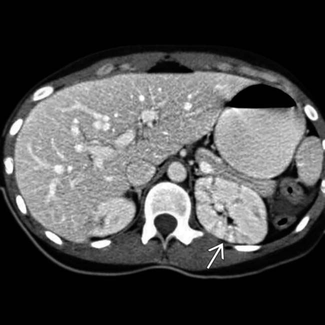
 representing acute ischemic injury.
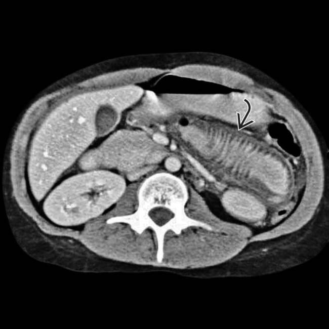
 , findings compatible with enteric ischemia. Rheumatoid vasculitis was subsequently confirmed. Findings that suggest bowel or renal ischemia in a young patient, as in this case, should raise suspicion for vasculitis.
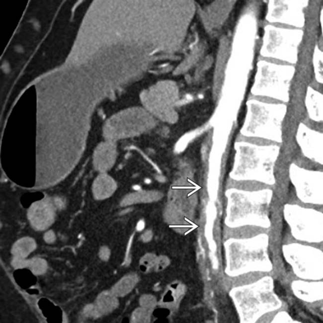
 .
.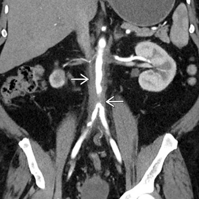
 of the abdominal aorta extending to involve the common iliac arteries. This is a common appearance for a large vessel vasculitis (giant cell vasculitis) with active inflammation.
 Classified by size of blood vessel involved into small vessel, medium vessel, and large vessel vasculitis


