Veins of the Spinal Cord and Spine
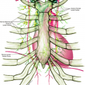
Inside and outside of the vertebral canal, along the entire length of the spinal cord, there are a number of venous plexuses, with free anastomoses with each other and connected with the intervertebral veins. Two groups of venous plexuses are found on the outside of the vertebral canal: the anterior group and the posterior group. The anterior group lies in front of the vertebral bodies and receives venous tributaries from other vertebral bodies and communicates with the basivertebral and intervertebral veins. It is most developed in the cervical region. The posterior group forms a network of venous plexuses mainly inside the spinal canal. There are three communicating valveless venous networks bearing a constant relationship to the vertebral bodies and intervertebral discs (Fig. 6.1). They are the intraosseous vertebral veins, the epidural venous plexus, and the paravertebral veins.
Veins of the Spine
Intraosseous Vertebral Veins
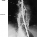
These veins drain the body of each vertebra. They empty into a venous sinus (the basivertebral vein) at the nutrient foramen of each vertebral body posteriorly (Fig. 6.2). These veins connect at each level with the second network, the epidural plexus.
Epidural Venous Plexus
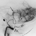
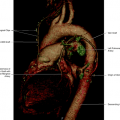
This plexus is composed of two vertical channels: the anterior internal vertebral veins that course the length of the spinal canal circling around the backs of the vertebral bodies and intervertebral discs, between the dura mater and bone. Both the left and right anterior internal vertebral veins have a lateral and a medial component. The lateral component is a single channel, whereas the medial component has a variable configuration and is a rather irregular group of vessels. The medial anterior internal vertebral veins are located close to the lateral anterior internal vertebral veins at all levels of the lumbar spine, except at the level L5–S1. At this level, the medial anterior internal vertebral veins leave the lateral anterior internal vertebral veins and lie close to the midline. The anterior internal vertebral veins are medial to the pedicles and bulge laterally as they cross the intervertebral disc spaces. The anterior internal vertebral veins communicate with the basivertebral veins through the nutrient foramina. There are segmental connections between the epidural venous plexus and the paravertebral veins at every level. At every intervertebral level there are two connecting veins on each side, named the supra- and infrapedicular veins (Figs. 6.2, 6.3, 6.4, 6.5).
There is also the posterior internal vertebral venous plexus, which is small and rudimentary, anastomosed to the anterior internal vertebral venous plexus through the lateral transverse branches. It is located on each side in front of the vertebral arches and ligamenta flava, having anastomoses with the posterior external plexuses by veins passing through and between the ligaments.
Related posts:
Stay updated, free articles. Join our Telegram channel

Full access? Get Clinical Tree


