• Villous adenoma is 1 histological type of adenomatous polyps (true neoplasms)
• Risk of cancer is related to tumor size, location, and proportion of villous change in adenoma
• Greater risk of carcinoma in villous tumors of stomach and duodenum than colon
Stomach: Carcinoma in 50% of lesions 2-4 cm and in 80% of lesions > 4 cm in size
Colon: Invasive carcinoma in up to 45% of cases
• CT: Large villous adenoma
Low-attenuation, minimally enhancing, irregular polypoid mass
Corrugated, feathery appearance due to trapping of enteric contrast
• Transrectal US; plus CT or MR for staging
TOP DIFFERENTIAL DIAGNOSES
• Colon carcinoma
• Fecal mass
PATHOLOGY
• Malignant potential: Lesions < 1 cm (5%), 1-2 cm (10%), > 2 cm (53%)
CLINICAL ISSUES
• Asymptomatic, diarrhea, pain, rectal bleeding, or melena
• Lesion closer to rectum: More likely to have diarrhea, electrolyte loss (hypokalemia and hyponatremia)
(Left) Graphic shows a polypoid mass in the rectosigmoid colon having a shaggy, nodular surface, sometimes likened to the surface of a cauliflower.
(Right) Single contrast barium enema shows a large rectal mass with a frond-like surface. Note the absence of a colonic obstruction, a typical feature of this soft and compressible tumor.
(Left) This 70-year-old man complained of frequent passage of watery stool, but had no symptoms of bowel obstruction. CT shows a large mass that fills the rectum. Note large vessels within and draining the mass.
(Right) Coronal CT reformation in the same case shows the huge size of the mass , but no definite signs of invasion through the rectal wall and no metastases. The resected villous adenoma had foci of frank carcinoma.
TERMINOLOGY
Synonyms
• Villous tumor
Definitions
• Adenomatous polyp that contains predominantly villous (“shaggy” surface) elements
IMAGING
General Features
• Best diagnostic clue
Polypoid lesion with nodular or frond-like surface on barium enema or CT colonography


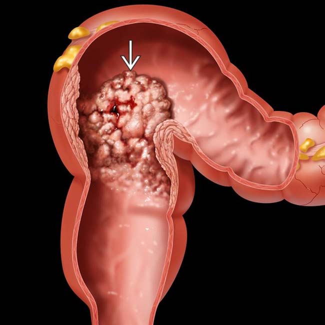
 in the rectosigmoid colon having a shaggy, nodular surface, sometimes likened to the surface of a cauliflower.
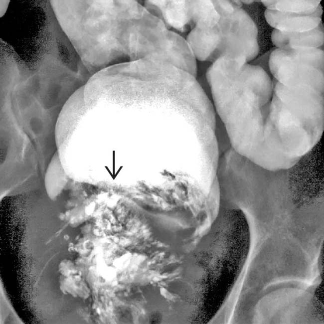
 with a frond-like surface. Note the absence of a colonic obstruction, a typical feature of this soft and compressible tumor.
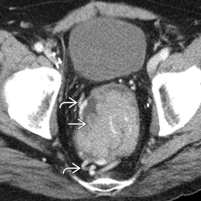
 that fills the rectum. Note large vessels
  within and draining the mass.
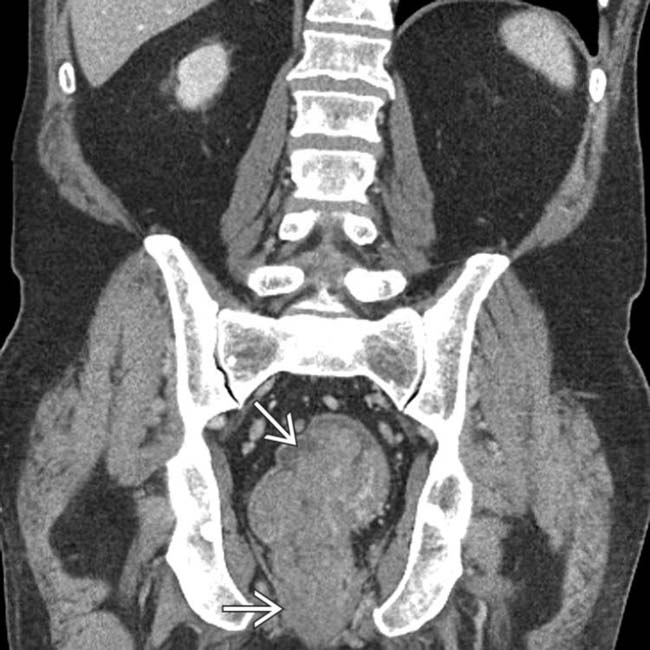
 , but no definite signs of invasion through the rectal wall and no metastases. The resected villous adenoma had foci of frank carcinoma.


