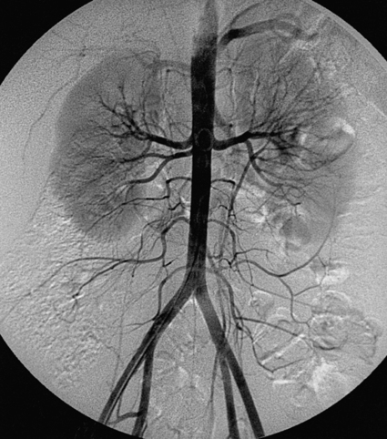
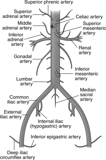
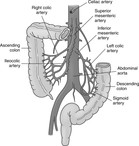
CHAPTER 14 After completing this chapter, the reader will be able to perform the following: The descending abdominal aorta provides the blood supply for the abdominal viscera. We have seen in Chapter 13 some of the major branches of the aorta. In this chapter we will discuss the vasculature of the gastrointestinal system, liver, spleen, and pancreas and the studies that relate to each of the respective organ systems. The blood supply for the abdominal viscera comes from the branches of the descending aorta (Fig. 14-1). These branches include the paired inferior phrenic, unpaired celiac trunk, superior mesenteric, paired middle adrenal, paired renal, paired gonadal, inferior mesenteric, and median sacral arteries in order of their origins from superior to inferior. Also between the levels of the paired renal arteries and the aortic bifurcation there are four paired lumbar arteries. We will confine our discussion of the vasculature to the following branches: celiac trunk and its branches and the superior and inferior mesenteric arteries (Fig. 14-2). The first branch of the common hepatic artery is the gastroduodenal artery. This vessel gives off the superior pancreaticoduodenal artery, which supplies the head of the pancreas with blood. Another branch continues as the right gastric epiploic artery to join with the left gastric epiploic artery. The common hepatic artery also gives rise to the right gastric artery, which runs toward the lesser curvature of the stomach and joins with the left gastric artery. At this point the hepatic artery is referred to as the hepatic artery proper. It courses upward to the liver and divides into the left and right hepatic arteries. The right hepatic artery usually gives off the cystic artery, which supplies the gallbladder with blood. The right and left hepatic arteries give off many branches and run in close proximity with branches from the hepatic portal vein. They empty into the hepatic sinusoids, which carry the blood to the central veins and out of the liver. Table 14-1 summarizes the various branches of the celiac trunk and the organ systems that are supplied by these vessels. TABLE 14-1 Summary of the Vasculature of the Celiac Trunk The superior mesenteric artery (Fig. 14-3) arises from the aorta approximately 1.5 cm below the celiac trunk. It lies at about the level of the first and second lumbar intervertebral space. It is responsible for providing the blood supply for the viscera from the middle portion of the duodenum to the transverse colon. The ileocolic branch of the superior mesenteric artery is the most inferior of the major branches. It courses toward the cecum and gives off an ascending branch, which joins with the descending branch of the right colic artery. The ileocolic branch terminates at the level of the ileocecal junction and divides into a number of smaller branches (Table 14-2). One of these branches, the appendicular artery, supplies the appendix with blood. TABLE 14-2 Summary of Vasculature of the Superior Mesenteric Artery The third major branch arising from the abdominal aorta is the inferior mesenteric artery (see Fig. 14-3). This vessel also provides a portion of the blood supply to the gastrointestinal system from the midtransverse colon to the rectum. It can be found on the ventral surface of the aorta at about the level of the third lumbar vertebra. The main trunk is longer than the superior mesenteric and is usually about 3 to 5 cm in length before it divides. The major branches from the vessel are the left colic artery, sigmoid arteries, and the superior rectal (hemorrhoidal) artery. The inferior mesenteric artery continues into the superior rectal (hemorrhoidal) arteries, which divide to form middle and inferior rectal branches. These vessels supply the rectum and the anal canal. Table 14-3 summarizes the vasculature of the inferior mesenteric artery. TABLE 14-3 Summary of Vasculature of the Inferior Mesenteric Branch of the Aorta
Visceral Angiography
 Describe the vascular anatomy of the abdominal viscera
 List the various procedures that can be performed in these areas
 List the indications and contraindications for angiography in these areas
 Explain vessel access for these procedures
 Describe the contrast agents used in visceral angiography
 List suggested patient positions for various visceral angiography studies
Anatomic Considerations
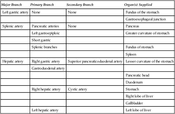
Celiac Trunk
Major Branch
Primary Branch
Secondary Branch
Organ(s) Supplied
Left gastric artery
None
None
Fundus of the stomach
Gastroesophageal junction
Splenic artery
Pancreatic arteries
None
Pancreas
Left gastroepiploic
Greater curvature of stomach
Short gastric
Splenic branches
Fundus of stomach
Spleen
Hepatic artery
Right gastric artery
Superior pancreaticoduodenal artery
Lesser curvature of the stomach
Gastroduodenal artery
Pancreatic head
Duodenum
Right hepatic artery
Cystic artery
Stomach
Right lobe of liver
Gallbladder
Left hepatic artery
Left lobe of liver
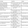
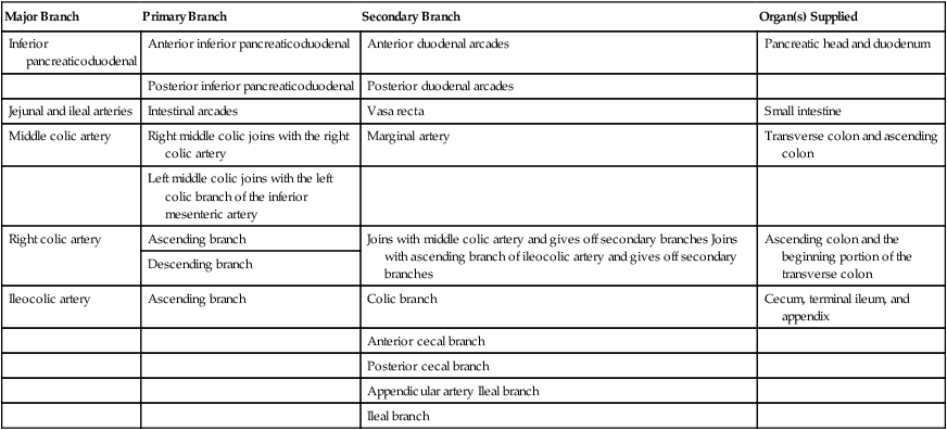
Superior Mesenteric Artery
Major Branch
Primary Branch
Secondary Branch
Organ(s) Supplied
Inferior pancreaticoduodenal
Anterior inferior pancreaticoduodenal
Anterior duodenal arcades
Pancreatic head and duodenum
Posterior inferior pancreaticoduodenal
Posterior duodenal arcades
Jejunal and ileal arteries
Intestinal arcades
Vasa recta
Small intestine
Middle colic artery
Right middle colic joins with the right colic artery
Marginal artery
Transverse colon and ascending colon
Left middle colic joins with the left colic branch of the inferior mesenteric artery
Right colic artery
Ascending branch
Joins with middle colic artery and gives off secondary branches Joins with ascending branch of ileocolic artery and gives off secondary branches
Ascending colon and the beginning portion of the transverse colon
Descending branch
Ileocolic artery
Ascending branch
Colic branch
Cecum, terminal ileum, and appendix
Anterior cecal branch
Posterior cecal branch
Appendicular artery Ileal branch
Ileal branch
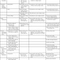
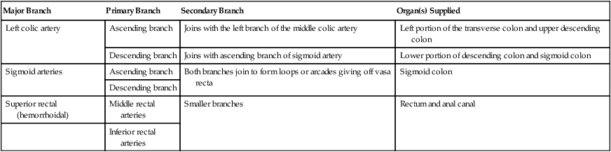
Inferior Mesenteric Artery
Major Branch
Primary Branch
Secondary Branch
Organ(s) Supplied
Left colic artery
Ascending branch
Joins with the left branch of the middle colic artery
Left portion of the transverse colon and upper descending colon
Descending branch
Joins with ascending branch of sigmoid artery
Lower portion of descending colon and sigmoid colon
Sigmoid arteries
Ascending branch
Both branches join to form loops or arcades giving off vasa recta
Sigmoid colon
Descending branch
Superior rectal (hemorrhoidal)
Middle rectal arteries
Smaller branches
Rectum and anal canal
Inferior rectal arteries
Visceral Angiography