Case presentation
A 14-year-old female is brought to the Emergency Department following a basketball game. She pivoted to take a shot and felt a tearing sensation, followed by a pop, then fell to the ground. She presents with an obvious right knee deformity. Her vital signs are normal for her age, and on examination, her knee is held in flexion at 30 degrees with a laterally palpable patella. She is grossly neurovascularly intact and there are no other injuries. Interestingly, the patient tells you she had the same injury twice in the past, most recently 5 months ago, involving the left knee, but never on the right. She is otherwise healthy.
Imaging considerations
Imaging is often employed in the evaluation of children with orthopedic complaints. Generally, imaging begins with plain radiography and progresses to other modalities based on history and physical examination. In the case of patellar dislocation, imaging may not be necessary prior to reduction unless there is concern for alternate diagnosis, as dislocation is often clinically apparent. Postreduction imaging is advised to evaluate for an osteochondral fracture, which is considered an indication for operative management ,
Plain radiography
The initial preferred imaging modality for a suspected patellar fracture is plain radiography. Anterior-posterior (AP), lateral, and oblique films should be requested, with the addition of a “sunrise view,” in the setting of concern for patellar injury. Prereduction imaging is likely unnecessary, as the diagnosis is usually clinically apparent, and the presence of fracture does not impact success of reduction. If prereduction radiographs are obtained, the dislocation can be readily identified; the patella tends to dislocate laterally. Postreduction imaging should be obtained on all patients to assess for fracture; however, plain radiography may have poor sensitivity for fracture. Multiple studies have revealed that fractures were present on arthroscopy in up to 70% of patients, which were not identified on plain radiography. Plain radiographs are also useful to evaluate patients who may have a predisposition to patellar dislocation, such as abnormal anatomy.
Magnetic resonance imaging (MRI)
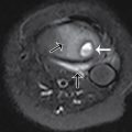
MRI is superior to plain radiography in identifying fracture and is the study of choice to detect injuries due to patellar dislocations that have spontaneously reduced. , , However, this is not a first-line imaging modality. MRI may be employed during follow-up evaluations, as it is useful for evaluating the articular cartilage, soft tissue injury, and medial patellofemoral ligament injury.
Ultrasound (US)
US is not a first-line imaging modality for patellar dislocation; however, it may be used to detect medial patellofemoral ligament injury during the postreduction follow-up period.
Computed tomography (CT)
CT is not a first-line imaging modality for diagnosing patellar dislocation. Use of this technology may be indicated in children with severe pain or associated complex fracture or when the diagnosis is uncertain. Primarily, CT is useful to assess the anterior tibial tubercle–trochlear groove distance, of which an abnormal measurement is an indication of patellar instability.
Imaging findings
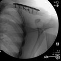
Prior to reduction, the patient had one-view imaging of the right knee. This image shows a laterally displaced patella without apparent fracture ( Fig. 66.1 ).

Case conclusion
The child’s clinical history, examination, and knee radiography were suggestive of patellar dislocation. She had a reduction performed immediately, which was successful. Postreduction imaging showed a reduced patella and no patellar fracture; there was remaining mild laxity ( Figs. 66.2–66.4 ). She was discharged in a knee immobilizer with planned outpatient orthopedic follow-up. At this evaluation, the patient and her family were presented with surgical and nonsurgical management options, given the multiple episodes of dislocation. Nonsurgical management was chosen, and the patient began a physical therapy regimen.


