Case presentation
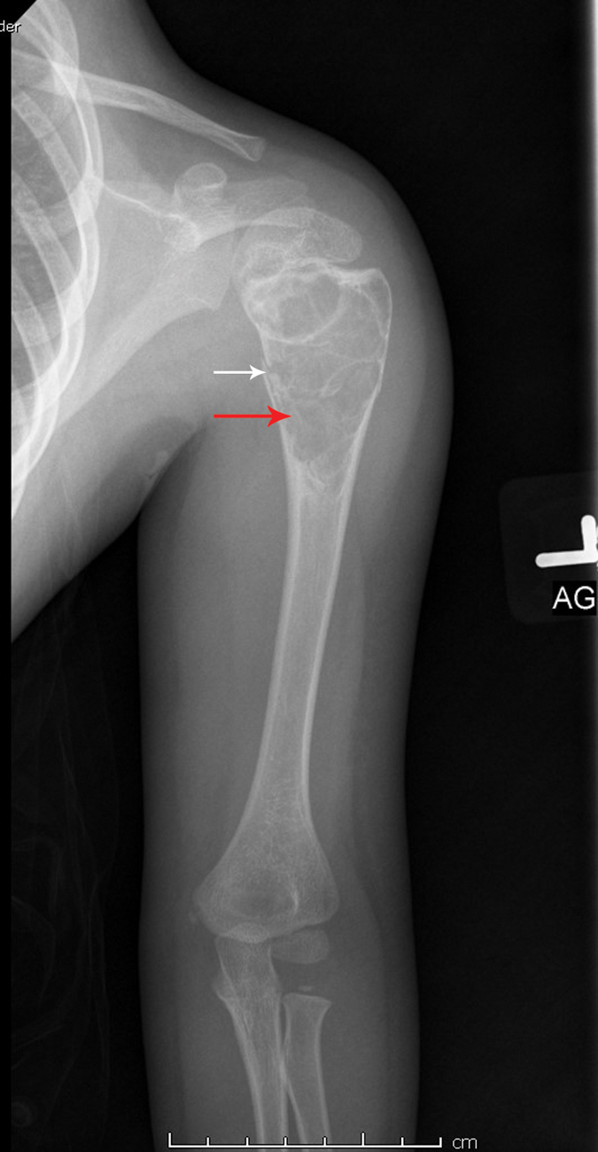
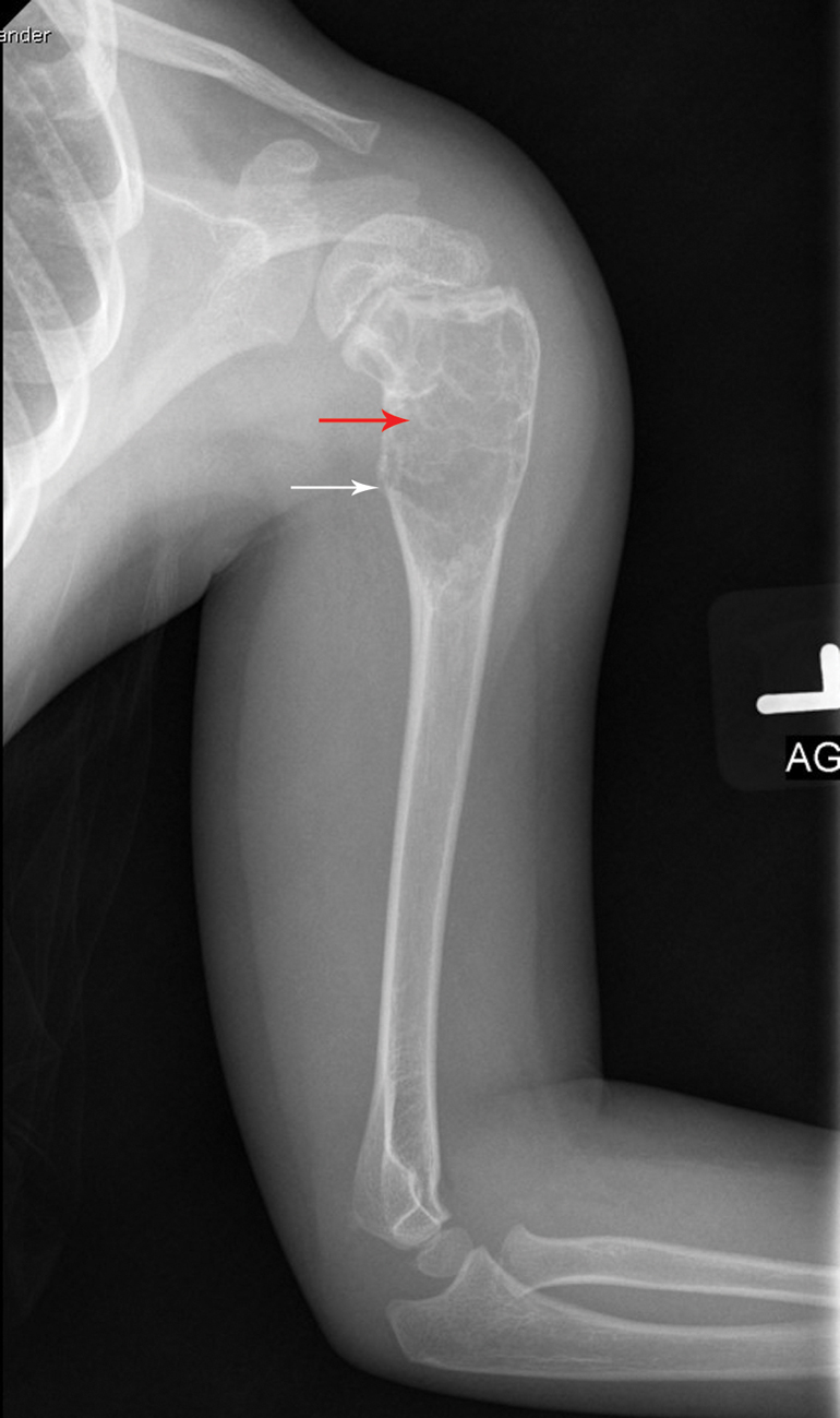
A 5-year-old male presents with left arm pain after a fall. He was playing soccer when he fell and landed on his left shoulder. He immediately complained of upper arm pain. His examination is significant for focal tenderness to the proximal left humerus with decreased range of motion when he attempts to raise the arm. There is mild edema. He is grossly neurovascularly intact. Imaging is obtained and a proximal humeral fracture is noted. However, the fracture is through a mildly expansile radiolucent lesion.
Prior to this trauma, the child had no symptoms, including fever, pain, or edema of the extremity. He also has no chronic medical conditions.
Imaging considerations
Plain radiography
As with most pediatric acute musculoskeletal trauma, plain radiography is the initial imaging modality of choice. When imaging the upper extremity in pediatric patients, two views (anteroposterior and lateral) of the involved extremity are appropriate. Consideration of imaging the joint above and below the suspected fracture area should be given, especially if there are historical or clinical indications of a second injury or where pain localization is difficult.
Computed tomography (CT)
CT, while excellent for evaluating fractures, is not indicated in the initial evaluation of suspected humeral fractures in children.
Magnetic resonance imaging (MRI)
MRI is not indicated in the initial imaging of musculoskeletal injuries in pediatric patients. This modality may be utilized if there are concerns for neurologic injury or compromise, although it is typically obtained after initial treatment and evaluation. MRI is often not readily available compared to plain radiography and CT. Younger children may require sedation.
Imaging findings
Two views (anteroposterior and lateral) of the injured arm are obtained. There is a nondisplaced fracture through a lucent lesion involving the proximal humeral metaphysis and diaphysis that is suggestive of a bone cyst ( Figs. 80.1 and 80.2 ). The radiographic appearance of the lesion is consistent with a simple bone cyst.