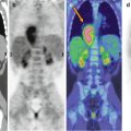
Fig. 16.1
A 3-year-old girl treated for Wilms’ tumor. Coronal (a–c) CT, PET, and PET/CT fusion images together with an axial (d) CT and PET/CT fusion image show an 18F-FDG-avid lesion at the upper pole of the right kidney, corresponding to disease recurrence
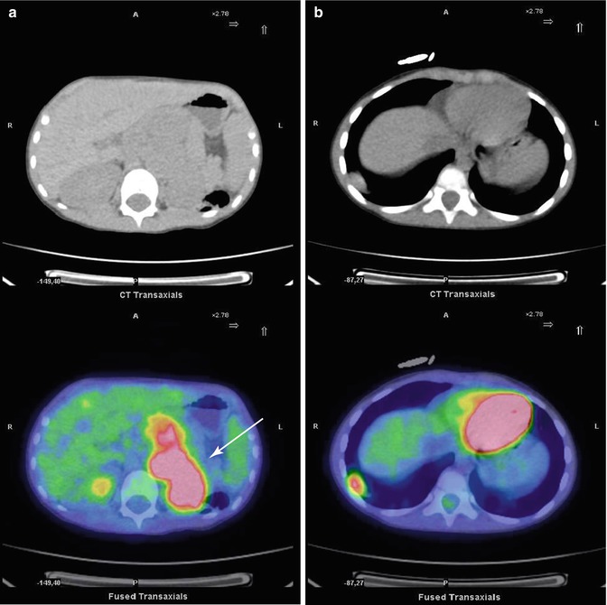
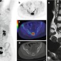
Fig. 16.2
A 3-year-old boy treated for Wilms’ tumor. Axial CT of the abdomen (a) and chest (b) and PET/CT fusion images show the extensive pathological involvement of the left kidney region (white arrow in a) and another accumulation in the basal segment of the lower right lung lobe
References
1.
2.
Charles AK, Vujanic GM, Berry PJ (1998) Renal tumours of childhood. Histopathology 32:293–309PubMedCrossRef
Stay updated, free articles. Join our Telegram channel

Full access? Get Clinical Tree


