X-ray Imaging: Mammography
Huimin Wu, PhD
LEARNING OBJECTIVES
1. Describe the main difference between the X-ray beams used for mammographic and radiographic imaging.
2. Name the commonly used materials for the anode target on a mammographic system.
3. Explain the role of the added beam filter in mammography.
4. Explain why molybdenum is never used as the filter when rhodium is the material for the anode target.
5. Name the main differences between screen-film cassettes used for mammography and those used for radiography.
6. Name the main reasons for breast compression in mammography.
7. List the methods commonly used for scatter reduction in mammography.
8. Explain how magnification mode works in mammography.
9. Explain the benefit of breast tomosynthesis in comparison with standard mammography.
OVERVIEW
Mammography is an X-ray imaging examination of breast tissues, primarily for detecting breast cancers. Breast cancers have both high morbidity and high mortality rates in the female population around the world. By estimate, one in eight women worldwide are affected by breast cancer. Early detection of breast cancer is considered crucial because treatment outcomes are typically better for breast cancers at earlier stages. Mammography plays a key role in the screening of breast cancer among asymptotic women, for the following reasons:
Mammography excels at detecting certain early signs of breast cancer, including microcalcifications, speculated masses, and distorted tissues.
Mammography demonstrates both high sensitivity and high specificity of cancer detection by clinical studies.
Mammography poses minimal risk to the subject with very low radiation dose.
Mammography is cost-effective and supports high throughput among a large population.
Mammography shares the same fundamental principles with radiography, but has its dedicated equipment and techniques to meet the unique challenges in breast imaging. For example, mammographic systems use X-ray beams of significantly lower energies than do standard X-ray equipment to increase the difference in attenuation between normal and cancerous tissues that would otherwise be too subtle for detection. Mammographic systems also use smaller focus spots and other measures to improve their spatial resolution for detecting microcalcifications. In addition, the level of radiation used by mammography is adjusted for each subject’s breasts and kept low to pose minimal risk to the subject.
X-RAY BEAM
Mammography requires the use of X-ray beams of lower energies compared to those of radiography, as a result of the inherently small difference in X-ray attenuation between normal and cancerous tissues (Figure 4.1). At X-ray energies of 40 keV or higher (the mean energy level standard radiography uses), the difference between the two types of tissues becomes too subtle to detect; so low-energy X-rays are preferred. On the other hand, X-ray energy cannot be too low because nearly all X-ray photons would be absorbed without reaching the detector, leading to unnecessary radiation dose to breast tissues. X-ray beam with photon energies around 20 keV is considered optimal for breast imaging, and this principle is integrated into the design of mammographic X-ray tubes.
The difference between mammography and radiography in their X-ray beam quality can be seen in differences in their half-value layers (HVLs). HVLs of an X-ray beam, defined as the thickness of a reference material (aluminum is most often used) needed to attenuate the beam intensity by half, are commonly used to characterize its beam quality. The higher the HVL, the higher mean energy the X-ray beam has. As an example (Figure 4.2), the HVL of a mammographic X-ray beam (assuming the use of molybdenum as anode target) is about 0.3 mm at 27 kVp, an order of magnitude smaller than that of a typical radiographic X-ray beam at 80 kVp.
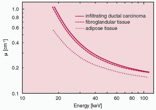 FIG. 4.1 • X-ray attenuation of breast tissues as a function of X-ray photon energy. Reprinted with permission from Andolina VF, Lillé SL. Mammographic Imaging. 3rd ed. Philadelphia, PA: Wolters Kluwer Health/Lippincott Williams & Wilkins; 2011. From Johns PC, Yaffe MJ. X-ray characterization of normal and neo-plastic breast tissues. Phys Med Biol. 1987;32(6):675-695. Copyright © 1987 Institute of Physics and Engineering in Medicine. Reproduced by permission of IOP Publishing. All rights reserved. |
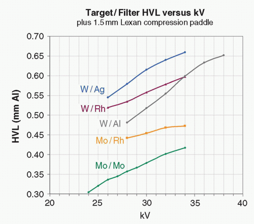 FIG. 4.2 • Half-value layers of mammographic X-ray beams. Reprinted with permission from Bushberg JT, Seibert JA, Leidholdt EM, Boone JM. Essential Physics of Medical Imaging. 3rd ed. Philadelphia, PA: Wolters Kluwer Health/Lippincott Williams & Wilkins; 2012. |
Table 4.1 MATERIAL PROPERTIES OF MOLYBDENUM, RHODIUM, AND TUNGSTEN, RELATED TO BEING ANODE TARGET OR BEAM FILTER FOR PRODUCING MAMMOGRAPHIC X-RAY BEAM. Those physical properties highly favorable for mammographic imaging are highlighted in bold; these include the energies of the characteristic x-rays from Mo and Rh and the high melting point of W. | ||||||||||||||||||||||||||||
|---|---|---|---|---|---|---|---|---|---|---|---|---|---|---|---|---|---|---|---|---|---|---|---|---|---|---|---|---|
|
Tube voltage
Low kVp is used by mammographic X-ray tubes to ensure that the average energy of produced X-ray beams is near 20 keV. For normal-sized breasts, tube voltage of 25 to 28 kVp is often used.
Cathode
A mammographic X-ray tube often has two filaments installed to produce two focal spot sizes; 0.3 and 0.1 mm are typical nominal values for the large and small focal spot sizes, respectively. In comparison, the focal spots on a radiographic X-ray tube are typically a few times larger. Tube currents available on a mammographic tube are also substantially lower than those by a radiographic tube, because of the overheating issues associated with smaller focal areas on the target for the interactions between electrons and the target.
Anode target
Molybdenum (Mo) and rhodium (Rh) have long been used as anode target materials in screen-film and digital mammography. Characteristic X-rays, highly dependent on the electron-shell structure of the anode material, account for a large fraction of the X-ray photons produced by the X-ray tube; Mo and Rh both have an electron structure that results in abundant X-ray photons near 20 keV (17.5 and 19.6 keV for Mo, and 20.2 and 22.7 keV for Rh), the desired energy range for breast imaging.
Tungsten (W) is another anode target material mostly used in digital mammography. With digital detectors, despite an unfavorable choice regarding characteristic X-ray production, W shows the following advantages as the target material: W provides higher X-ray production efficiency, because of its higher atomic number, and much improved heat loading, because of its higher melting point, compared to Mo and Rh; in addition, wider dynamic range and postprocessing capabilities of digital detectors relax the requirement for X-ray beam energy, making characteristic radiation from Mo or Rh not as crucial as it is in screen-film mammography.
Table 4.1 shows a comparison of the three anode target materials in their physical properties related to mammography.
Beam filter
Both the tube port, as part of the inherent filtration, and the added beam filters play important roles in shaping the X-ray energy spectrum produced by a mammographic X-ray tube. Beryllium (Be) is typically used for the tube port where the beam exits the tube. Be has a low atomic number of 4 and is strong enough to be made thin. The thin tube port window made by Be absorbs low-energy X-ray photons that are unable to penetrate breasts and lets through those with higher energies.
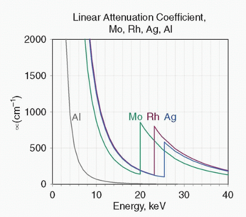 FIG. 4.3 • The attenuation characteristics of filter materials used on mammographic systems, including Mo, Rh, Ag, and Al. Mo, Rh, and Ag all have K-edges between 20 and 30 keV, useful for removing photons of high energy that could compromise tissue contrast otherwise. Reprinted with permission from Bushberg JT, Seibert JA, Leidholdt EM, Boone JM. Essential Physics of Medical Imaging. 3rd ed. Philadelphia, PA: Wolters Kluwer Health/Lippincott Williams & Wilkins; 2012. |
Added X-ray tube filtration further shapes the energy spectrum of the X-ray beam by selectively removing undesired X-ray photons with either too high or too low energies. It is often accomplished using materials that have K-edge energies that are slightly above the upper bound of the optimal energies for breast imaging. The K-edge of an element is the energy level at which X-ray photons have just enough energy to knock out atomic electrons on the K-orbit and a substantial increase in X-ray absorption results. Mo, Rh, and Ag are typical elements with suitable K-edge energies for providing added beam filtration for mammography (Figure 4.3). The added filter heavily absorbs both low-energy photons (<15 keV), due to high photoelectric absorption, and high energy ones (above the K-edge), due to dramatically increased absorption beyond the K-edge. The result is selective transmission of X-rays in a narrow band of energies from about 15 keV up to the K-absorption edge of the filter.
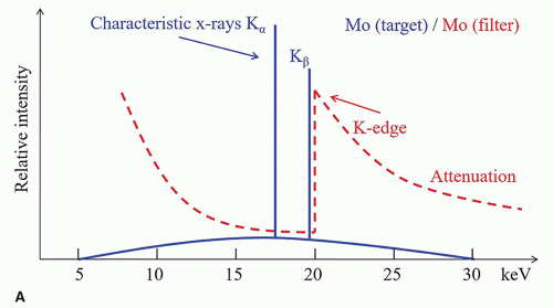 FIG. 4.4 • A-D: Choice of appropriate target-filter combinations when Mo and Rh are used as target and filter. Mo/Mo, Mo/Rh, and Rh/Rh all provide X-ray spectra suitable for mammography with mean energies around 20 keV; the combinations with Rh as filter have higher average energy because the spectra are cut off at higher energy by the K-edge of Rh. Rh/Mo would not work well for mammography because Rh-produced characteristic X-rays are removed by the K-edge absorption by the Mo filter. |
Target-filter combination
The selection of the appropriate combination of target-filter materials is a nontrivial matter in mammographic tube design. When Mo or Rh is used as the target material, because of the abundant characteristic X-rays in the produced spectrum, the K-edge of the added filter needs to be slightly above the energies of those characteristic X-rays so as not to absorb these useful photons (Figure 4.4). If Mo is used as the target material, both Mo and Rh can be used as the filter materials. If Rh is used as the target material, then Rh itself is an acceptable filter material, but Mo would not be a good candidate because its K-edge is lower than the energies of characteristic X-rays produced by an Rh target.
When W is used as the target material, practical choices of filter materials include Rh, Ag, and Al. Because of its large atomic number, W produces a smaller fraction of characteristic X-rays than do Mo and Rh as a target, and these photons are at higher energies than the K-absorption edges of the filter materials listed. Rh is routinely used for imaging of normal-sized or smaller breasts. The energy band of transmitted X-rays for Ag is shifted toward the higher end compared to that for Rh, so Ag is often the choice when imaging larger than normal breasts. Figure 4.5 shows X-ray spectra of the target-filter combinations of W/Rh and W/Ag. Al is mostly used for tomosynthesis acquisition, a mode of multiview acquisition for producing a 3D-like image set. Al does not have a sufficiently high K-edge to reject higher energy photons, so it primarily serves to remove low-energy ones for dose savings. The rejection of higher energy photons is not a strong need in tomosynthesis for image contrast considerations, because of the improvement in image contrast that comes with the reconstruction process in tomosynthesis.
Related posts:

Stay updated, free articles. Join our Telegram channel

Full access? Get Clinical Tree


