Discrete intramural nodules appear T2 hyperintense and hypointense on T1WI and T1WI C+
Thickened gallbladder wall and intramural nodules may show signal drop-out on out-of-phase images
Thickened wall often demonstrates delayed enhancement on T1WI C+
• US: Hypoechoic nodules or bands within thickened GB wall
Gallstones, sludge, echogenic intraluminal debris
PATHOLOGY
• Mucosal ulceration or rupture of Rokitansky-Aschoff sinuses → extravasation of bile into GB wall → phagocytosis of bile lipids → inflammation and xanthoma cell formation
• Gallstones always present and may play causative role
CLINICAL ISSUES
• Symptoms most often similar to acute cholecystitis, but can be chronic and mimic malignancy
• More common in females during 6th or 7th decade of life
• Only definitive treatment is cholecystectomy
(Left) Resected GB shows marked wall thickening and an intramural abscess . A portion of the adjacent liver was resected because of the high intraoperative suspicion of GB cancer. Pathology revealed xanthogranulomatous cholecystitis (XGC).
(Right) Axial CECT shows a thickened wall of the gallbladder, especially the fundus , with an indistinct border with the liver. While the appearance was concerning for gallbladder cancer, this was found to be XGC at cholecystectomy.
(Left) Axial CECT of a patient with RUQ pain shows marked irregular GB wall thickening , intramural low attenuation , and several pericholecystic collections . Low-attenuation intramural nodules in XGC are due to either abscesses or xanthogranulomas.
(Right) Axial NECT in a patient with chronic abdominal pain shows a distended, thick-walled gallbladder exhibiting indistinct margins with the liver . While this was worrisome for carcinoma, XGC was confirmed at surgery.
 Pericholecystic fluid and inflammatory change ± fistulous tracts, abscesses, contained perforation, etc.


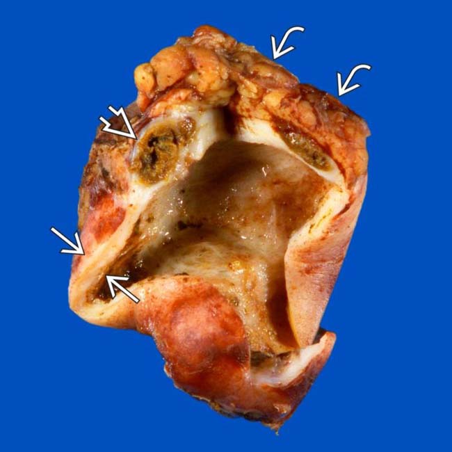
 and an intramural abscess
  . A portion of the adjacent liver
  was resected because of the high intraoperative suspicion of GB cancer. Pathology revealed xanthogranulomatous cholecystitis (XGC).
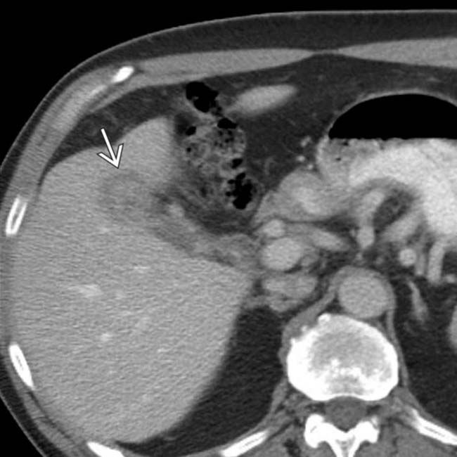
 , with an indistinct border with the liver. While the appearance was concerning for gallbladder cancer, this was found to be XGC at cholecystectomy.
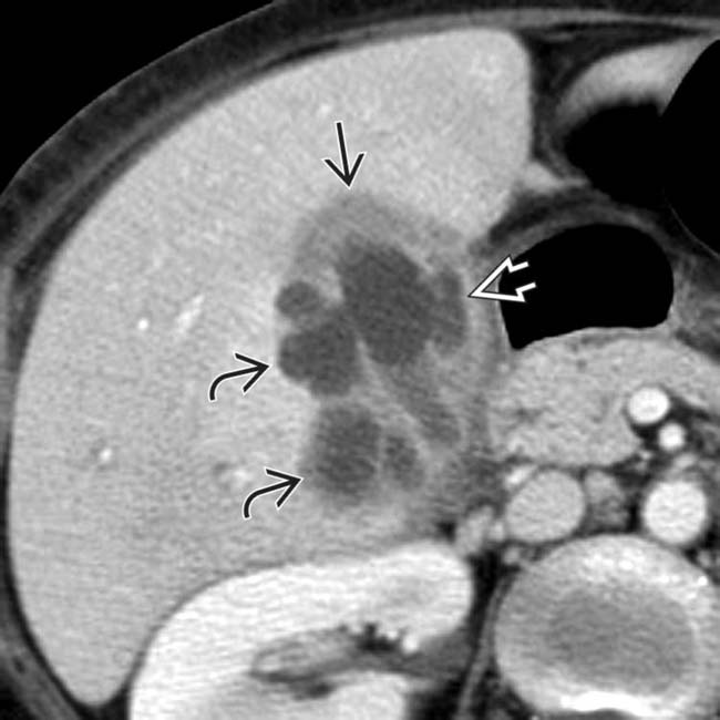
 , intramural low attenuation
  , and several pericholecystic collections
  . Low-attenuation intramural nodules in XGC are due to either abscesses or xanthogranulomas.
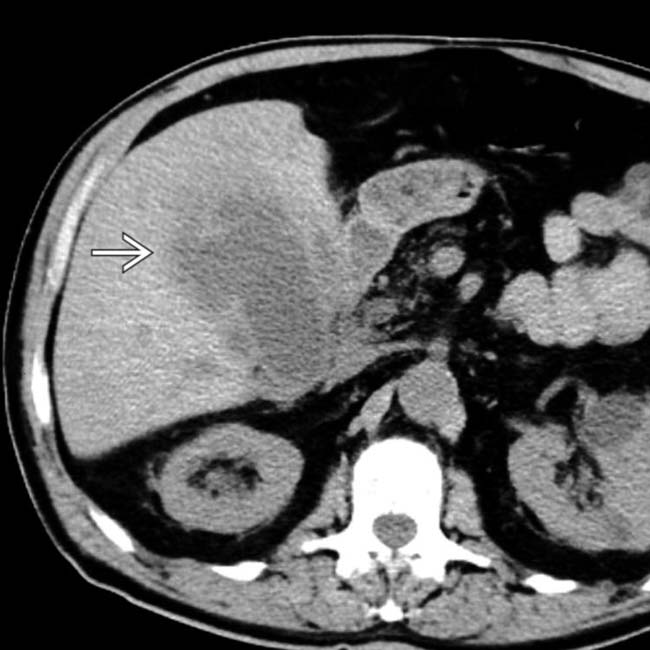
 . While this was worrisome for carcinoma, XGC was confirmed at surgery.


 Low-attenuation intramural nodules and bands corresponding to foamy cell infiltrate and areas of necrosis/abscess
 Pericholecystic fluid, inflammatory change, and induration ± fistulous tracts, abscesses, contained perforation, etc.


