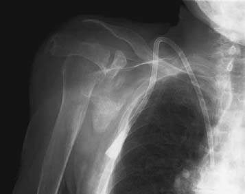
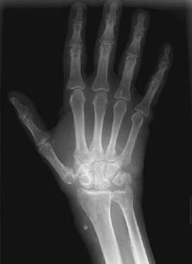
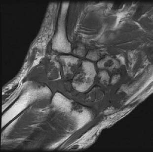
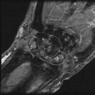
CASE 118 Peter L. Munk and Anthony G. Ryan A 70-year-old woman presented with complaints of diffuse pain throughout her right upper limb, with more focal pain at the wrist and shoulder. On examination, soft-tissue swelling was evident on the dorsum of the hand. Figure 118A Figure 118B Figure 118C Figure 118D An anteroposterior (AP) shoulder radiograph (Fig. 118A) shows a right internal jugular hemodialysis catheter in situ. There is diffuse osteoporosis. The clavicle is discontinuous laterally, the inferior portion of which is tapered. The lateral portion is projected anterior to the head of the humerus, which is deformed and subluxated in relation to the glenoid fossa. An AP radiograph of the right hand and wrist (Fig. 118B) shows diffuse soft-tissue swelling overlying the dorsum of the hand and distal wrist. Multiple erosions are evident, for example, at the base of the fifth metacarpal and distal ulna, as is deformity of the proximal carpal row bones and trapezium, the latter associated with subluxation of the first carpometacarpal joint. Calcification is evident within the ulnar artery, as is a phlebolith in a previous fistula site on the radial aspect of the wrist. Cystic change is evident at the base of the fifth metacarpal. A coronal T1-weighted image (Fig. 118C) shows low signal intensity material essentially filling the joint space and eroding into the carpal bones, almost replacing some of the latter, particularly the proximal carpal row. Only a very small portion of the trapezium remains. Erosions secondary to the encroaching material are evident within the distal radius and ulna, as well as at the base of the second metacarpal. A corresponding postgadolinium image (Fig. 118D) shows that the eroding material enhances moderately. Dialysis-induced amyloid arthropathy. Despite the widespread use of renal transplantation, a larger number of patients are currently undergoing hemodialysis than at any other time in history. These patients often will develop arthritic and periarthritic changes. Many of these changes are due to osteodystrophy and secondary hyperparathyroidism, both of which are dealt with separately in other chapters. Many of the changes are, however, due to deposition of amyloid. Beta2-microglobulin is a protein polysaccharide (frequently consisting of immunoglobulin light chains) that, the presence of a normally functioning glomerulus, is freely filtered from serum. In patients without glomerular function, this protein accumulates in serum and frequently reaches levels 40 to 50 times that normally encountered. This is not reduced by hemodialysis, as the cellulose-based hemodialysis membranes cannot filter beta2-microglobulin. This results in a tendency to systemic deposition via the capillaries of a variety of different tissues.
Amyloid Arthropathy Secondary to Renal Failure
Clinical Presentation
Radiologic Findings
Diagnosis
Differential Diagnosis
Discussion
Background
Etiology
Related posts:
Stay updated, free articles. Join our Telegram channel

Full access? Get Clinical Tree


