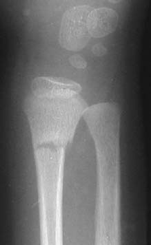
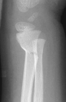
CASE 100 Peter L. Munk, Anthony G.Ryan, and Laurel O. Marchinkow A 5-year-old boy, having fallen from a chair, presented with pain, swelling, and reduced movement of his right arm. Figure 100A Figure 100B Anteroposterior (AP) (Fig. 100A) and lateral (Fig. 100B) images of the wrist show an incomplete fracture of the distal radius affecting the ulnar and volar aspects of the bone; however, the dorsal and radial aspects of the cortex are not breached. Greenstick fracture of the distal radius. None. The combination of Salter-Harris and greenstick fractures accounts for the vast majority of fractures seen in the pediatric population. The pediatric skeleton is far more plastic than the adult, frequently resulting in incomplete fractures due to the deformability of the immature bones. Several types of incomplete fractures are commonly encountered. Notable are two principal types of fractures that typically affect long bones, particularly in the upper extremity: greenstick and torus. These fractures are essentially confined to the pediatric population, and a typical history of fall followed by immediate pain and swelling is usually given. A greenstick fracture occurs due to a bending force on the bone, resulting in fracturing of the cortex on one side of the bone but not the other (Fig. 100C). The convex side of the stressed bone shows a radiolucent fracture line that does not extend to the concave side (Figs. 100A, 100B
Greenstick Torus Fracture
Clinical Presentation
Radiologic Findings
Diagnosis
Differential Diagnosis
Discussion
Background
Etiology
Pathophysiology
Clinical Findings
Complications
Imaging Findings
RADIOGRAPHY
Greenstick fracture
![]()
Stay updated, free articles. Join our Telegram channel

Full access? Get Clinical Tree


