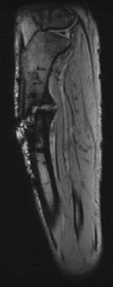
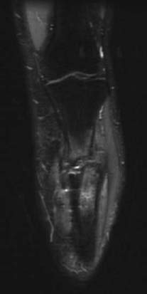
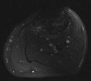
CASE 101 Hema N.Choudur, Anthony G. Ryan, and Peter L. Munk A young man had had a motor vehicle accident several months prior to presenting. He had had a fracture of the right tibia, which had been casted; however, follow-up radiographs did not show any healing. Because there was severe pain at that site, an MRI was recommended to visualize any bony union and to exclude infection. Figure 101A Figure 101B Figure 101C The MRI (Fig. 101A, sagittal MPGR; Fig. 101B, coronal FSTIR; and Fig. 101C, axial T1-weighted fatsaturated postgadolinium) reveals no osseous union between the fractured fragments. Although edema is evident in the adjacent marrow, there is no abnormal enhancement postgadolinium to suggest infection. Nonunion. The repair of a fracture is a continuous process, with the fracture tender and slightly mobile in the early stages becoming immobile and nontender in the later stages, noticeably seen on plain radiographs as bony fusion as against callus formation in the early stages.
Fracture Nonunion
Clinical Presentation
Radiologic Findings
Diagnosis
Differential Diagnosis
Discussion
Related posts:
Stay updated, free articles. Join our Telegram channel

Full access? Get Clinical Tree


