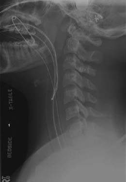
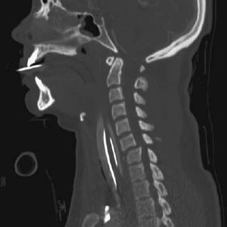
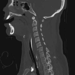
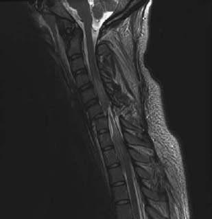
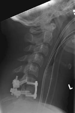
CASE 104 Hema N. Choudur, Anthony G. Ryan, and Peter L. Munk Following a diving injury in shallow waters, a young man was brought to the emergency department with quadriplegia. Figure 104A Figure 104B The lateral radiograph (Fig. 104A) reveals an anterosuperior C7 vertebral fracture with mild displacement of the fractured fragment. Also noted is the fracture of the superior articular process of the same vertebra. Sagittal reformats from the subsequent CT (Figs. 104B, 104C) show the C7 fracture fragment and C6 over C7 anterior subluxation causing spinal canal narrowing at the same level. A T2-weighted sagittal image from the subsequent MRI (Fig. 104D) shows the C6 on C7 anterior subluxation with a tear of the posterior longitudinal ligament and buckling of the anterior longitudinal ligament. A hematoma interposed between the anterior longitudinal ligament and the vertebral body is evident. Clearly depicted is the transection of the cord at the same level, with post-traumatic high signal intensity within the cord above the level of injury. A postfixation cervical spine lateral view (Fig. 104E) shows stabilization of the vertebrae at the level of the injury. Figure 104D Figure 104E Flexion teardrop fracture with associated cord transection. Wedge compression fractures are usually stable injuries. The posterior cortex is intact with no ligamentous injury. These fractures were originally described by Schneider and Cann as “teardrop” fractures. Teardrop fractures represent 15% of traumatic cervical spine injuries. Injuries can be severe, with cord involvement and quadriplegia in 60% of cases. These fractures are very unstable. The kyphosis can progress with neurologic deterioration. Stabilization is by operative means.
Teardrop Fracture
Clinical Presentation
Radiologic Findings
Diagnosis
Differential Diagnosis
Discussion
Background
Stay updated, free articles. Join our Telegram channel

Full access? Get Clinical Tree


