Midsagittal non-contrast T1WI of the brain through the corpus callosum and pituitary gland.
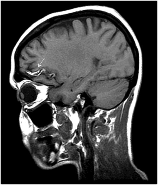
Parasagittal non-contrast T1WI of the brain through region of left frontal operculum.
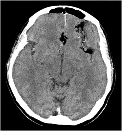
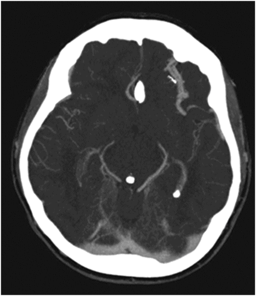
Axial non-contrast CT at the level of ambient cistern and sylvian fissures.
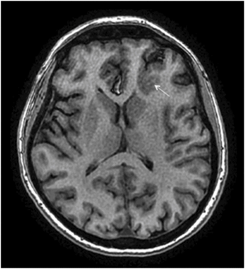
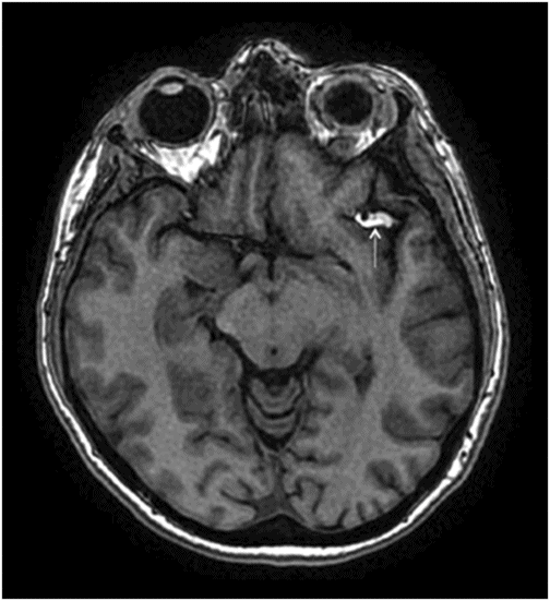
Axial non-contrast T1WI of the brain at the level of lateral ventricles and inferior frontal gyrus.
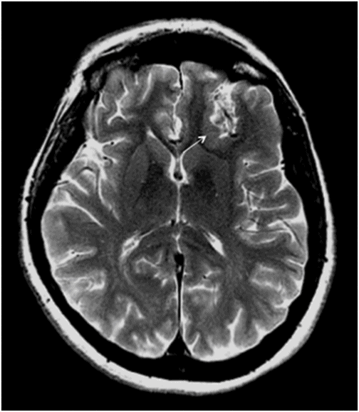
Axial T2WI of the brain at the level of lateral ventricles and inferior frontal gyrus.
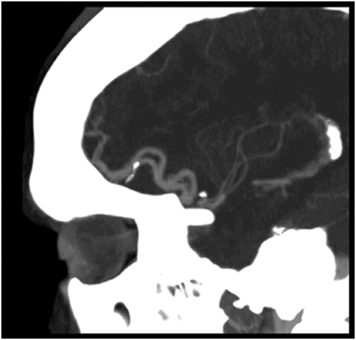
Parasagittal reformatted CTA of circle of Willis through region of left frontal operculum.
Interhemispheric and Pericallosal Lipoma with Cortical Dysplasia
Primary Diagnosis
Interhemispheric and pericallosal lipoma with cortical dysplasia
Differential Diagnoses
Dermoid
Osteolipoma
Encephalocraniocutaneous lipomatosis
Imaging Findings
Fig. 109.1: Sagittal T1WI demonstrated a curvilinear pericallosal-interhemispheric lipoma, but otherwise normal-appearing morphology of corpus callosum. Fig. 109.2: Parasagittal T1WI showed extension of the lipoma into the left inferior frontal sulcus and pars opercularis (arrow). Fig. 109.3: Axial T1WI through the level of the cerebral peduncles demonstrated lesion extension into the proximal sylvian fissure (arrow). Fig. 109.4: Axial CT demonstrated fat density in the left inferior frontal sulcus and interhemispheric fissure (arrows). Fig. 109.5: Axial T1WI and Fig. 109.6: Axial T2WI demonstrated a dysplastic left inferior frontal gyrus (arrows). Fig. 109.7: Axial CT angiography and Fig. 109.8: Oblique sagittal images demonstrated dilated, tortuous arteries.
Discussion
The T1 hyperintense signal extending along the interhemispheric fissure and pericallosal location, inferior frontal sulcus, frontal operculum, and proximal sylvian fissure with fat attenuation density on CT confirms the diagnosis of lipoma.
The major differential diagnosis to consider is a dermoid cyst, which also shows evidence of fat density and is usually located in the midline. They are non-enhancing, lobulated, and well defined, unless ruptured, when they present with fat globules in the subarachnoid space. Dermoids tend to have variable solid-cystic components. However, lipomas often have near homogeneous fat density artifact. Osteolipomas are lesions that are peripherally calcified with central fatty component and can be differentiated from lipoma.
Encephalocraniocutaneous lipomatosis (ECCL) is a complex syndrome with leptomeningeal lipomatosis, lipomas involving the skull, heart, and eyes, brain malformations, and mental retardation. Absence of multiple lipomas and mental retardation in this patient case excludes ECCL. Several intracranial tumors can have lipomatous differentiation/transformation such as neuroectodermal tumors, glial neoplasms of brain and spinal cord, meningiomas, and cerebellar liponeurocytomas.
Intracranial lipomas (also known as lipomatous hamartomas) are rare congenital brain malformations, with an estimated incidence of 0.1–0.4% of all brain tumors. Clinically, they can be difficult to diagnose, as they are often asymptomatic. Occasionally patients with intracranial lipomas can present with seizures (up to 50% of patients) or mental disorders. They have also been described in association with a rare inherited disorder, familial multiple lipomatosis (FML).
Lipomas typically occur in the midline and over 80% are found supratentorially. Approximately 20% occur infratentorially, typically in the cerebellopontine angle, but can also be seen in the jugular foramen and foramen magnum. They are predominantly found above the corpus callosum (~30–40%) and termed interhemispheric lipomas. These may extend into the lateral ventricles and choroid plexus. Frequently, they are seen in the pericallosal location. They can also be seen in the pineal region (20–25%), attached to the tectum suprasellar region (15–20%), or attached to the tuber cinereum. Small lipomas involving the sylvian fissures, middle cranial fossa, and insular regions are rare and account for 3% of the intracranial lipomas.
The pathogenesis of intracranial lipomas is not clearly established; however, the leading theory suggests they result from abnormal persistence and abnormal differentiation of the meninx primitiva, the undifferentiated mesenchymal precursor of the leptomeninges, during the development of subarachnoid cisterns. Other theories suggest the lesions represent hypertrophy of preexisting meningeal adipose tissue, metaplasia of meningeal connective tissues, or a defective junction of the cutaneous ectoderm recovering the mesoderm occurs during neural tube development.
More than 50% of lipomas occur in conjunction with brain malformations of varying degrees. They can contribute to the development of focal cortical dysplasia as demonstrated in our case, because of the physical cortical interruption and focal perfusion insufficiency. Lipomas are associated with various CNS anomalies such as agenesis/dysgenesis of the corpus callosum, absence of septum pellucidum, cranium bifidum, spina bifida, encephaloceles, and myelomeningoceles. Pachygyria, polymicrogyria, and gray matter heterotopia have all been described in lipomas. Hypervascularization surrounding the lipoma can occur with adjacent venous angiomas, saccular aneurysms, dilated and tortuous feeding arteries, and abnormal arterial branches.
On imaging, lipomas are well-delineated lobulated extra-axial/pial-based masses with fat attenuation. Interhemispheric lesions can present as two types – curvilinear (thin lipoma which curves around the corpus callosum) or tubulonodular (bulky mass often seen in association with calcifications and usually associated with corpus callosum agenesis). On MR imaging they are hyperintense on T1WI and appear hypointense on T1- and T2-weighted fat-suppression techniques. Adipose tissue lesions also feature chemical shift artifact.
Patients with hemispheric lipomas often respond to medical management. Given the paucity of cases, their response to surgical management cannot be accurately evaluated.
Stay updated, free articles. Join our Telegram channel

Full access? Get Clinical Tree


