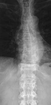
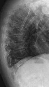
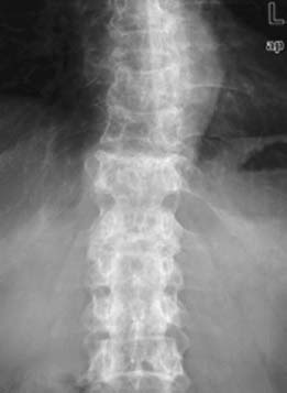
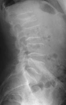
CASE 109 Hema N. Choudur, Anthony G. Ryan, and Peter L. Munk An elderly woman complained of long-standing back pain with no history of trauma. On questioning, she admitted having widespread bony pain. Figure 109A Figure 109B Figure 109C Figure 109D Plain radiographs of the dorsal and lumbar spine anteroposterior (AP) and lateral views (Figs. 109A–109D), show mid-dorsal and L2 compression fractures with no paravertebral soft tissue and normal adjacent disk spaces. There was a generalized decrease in bone density. Compression fractures. The most common cause of compression fracture is osteoporosis. These fractures can occur without trauma and remain silent. Progressive kyphosis may be the only indication of a compressed vertebra. Some patients have intense pain at the site of the vertebral compression fracture. Most commonly, these patients are treated conservatively; however, in a significant proportion, back pain persists, and the prolonged immobilization places them at risk for complications, such as pneumonia, urosepsis, and deep venous thrombosis. These patients may now be treated with vertebroplasty or kyphoplasty, which alleviates the pain and permits return to full mobilization in over 80% of cases. Most nontraumatic compression fractures are osteoporotic. One third are seen in the lumbar vertebrae, a third in the thoracolumbar vertebrae, and a third in the thoracic vertebrae. Seventy-five percent of women older than 65 years with scoliosis have at least one osteoporotic wedge fracture. Other causes include trauma, malignancy, infection, and hemangiomas. Traumatic fractures are frequent in the dorsolumbar junction.
Vertebral Compression Fractures
Clinical Presentation


Radiologic Findings
Diagnosis
Differential Diagnosis
Discussion
Background
Etiology
Pathophysiology
Stay updated, free articles. Join our Telegram channel

Full access? Get Clinical Tree


