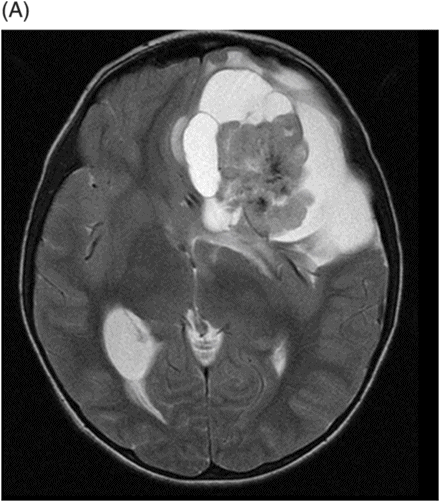
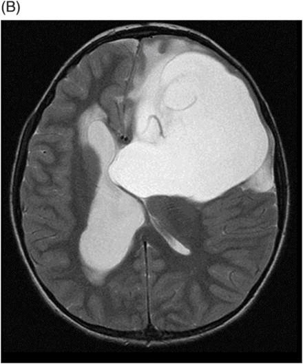
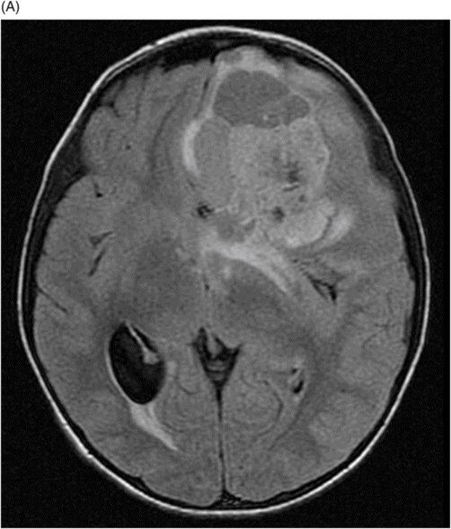
(A–B) Axial T2WI through the frontal lobe.
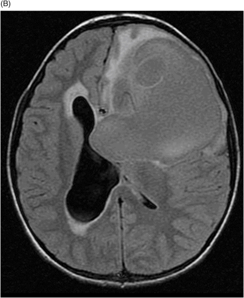
Axial GRE-T2* WI sequence through the lateral ventricles.
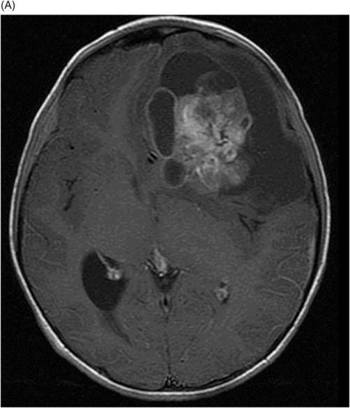
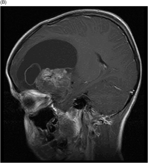
(A) Axial T1WI and (B) Sagittal T1WI postgadolinium through the lesion in the left frontal lobe.
Supratentorial Ependymoma
Primary Diagnosis
Supratentorial ependymoma
Differential Diagnoses
Atypical teratoid/rhabdoid tumor
Primitive neuroectodermal tumor
Teratoma
Choroid plexus carcinoma
Imaging Findings
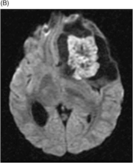
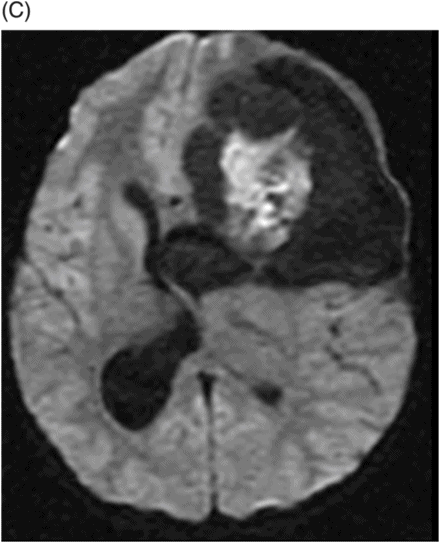
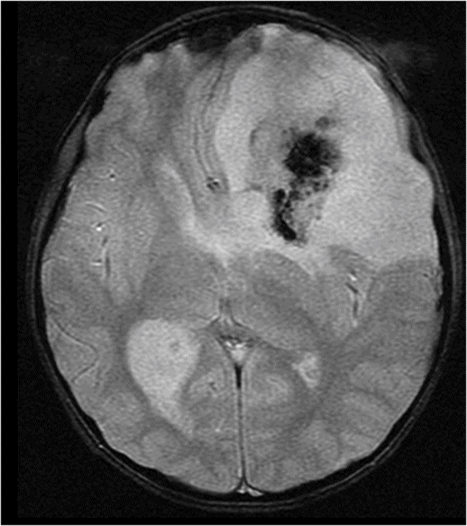
Fig. 110.1: (A) Sagittal T1WI showed a large, subcortical, solid-cystic mass in the left frontal lobe that is iso- to hypointense. (B–C) Axial DW MR images showed restricted diffusion in the solid component of the mass. Fig. 110.2: (A–B) Axial T2W images demonstrated a large frontal mass, with low signal on T2. Fig. 110.3: (A–B) Axial FLAIR images showed a moderately hyperintense tumor with central areas of high signal intensity due to necrosis. Fig. 110.4: Axial GRE-T2* WI sequence showed hypointense areas in the solid component representing hemorrhage or calcifications. Fig. 110.5: (A) Axial T1W and (B) Sagittal T1W postgadolinium images demonstrated a moderate enhancement of the tumor with non-enhancing areas of necrosis.


Stay updated, free articles. Join our Telegram channel

Full access? Get Clinical Tree


