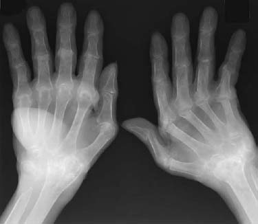
CASE 111 Peter L. Munk and Anthony G. Ryan A 56-year-old woman presented with chronically painful and stiff hands and wrists. The stiffness was maximal on rising and improved slightly after an hour or so. Marked soft-tissue swelling was evident on examination of the dorsum of the right wrist and around the left second and third metacarpophalangeal (MCP) joints. Figure 111A Radiograph of both hands (Fig. 111A) shows tapering absence of both distal ulnae and almost complete absence of the carpal bones of each wrist. The radius appears to articulate directly with the metacarpals bilaterally. Almost complete erosion of the metacarpal heads resulting in ulnar deviation and partial subluxation of the MCP joints is evident on the left. Less advanced erosive change at the MCP joints is evident on the right. Further erosive changes are evident at the heads of the proximal phalanges, giving rise to subluxation of the proximal interphalangeal (PIP) joints bilaterally. Massive soft-tissue swelling is evident projected through the ulnar aspect of the dorsum of the hand on the right. Less prominent soft-tissue swelling is evident over the second and third MCPs on the right and further soft-tissue swelling overlying the second to fourth MCPs on the left. The bones are diffusely osteopenic. Severe advanced rheumatoid arthritis. Rheumatoid arthritis and juvenile rheumatoid arthritis appear to be two distinctly separate diseases, although there may be some cases that occur in children that more closely resemble the adult form. The etiology of rheumatoid arthritis remains controversial. Autoimmune phenomena have often been used to explain this disease process, although others advocate that a close relationship with an infectious agent may exist. An increased incidence of rheumatoid arthritis has been demonstrated in patients with human leukocyte antigen (HLA) DR4. An increased incidence postpartum has been identified. A diffuse inflammatory synovitis results in inflammatory cells and macrophages replacing normal synovial tissue, occasionally replacing the complete synovial membrane. The infiltrated/abnormal “synovium” is often referred to as pannus, which is typically highly vascular and can become markedly hypertrophic, filling the joint completely with very little of the joint space occupied by fluid. Pannus is also aggressively destructive, in part by preventing synovial fluid from nourishing cartilage, and also by the release of a variety of lysosomal enzymes that destroy cartilage and adjacent soft tissues and can even result in extensive osseous destruction. As the disease continues to progress, and more and more pannus is elaborated, narrowing of the joint space occurs, especially as pannus extends across the joint, destroying cartilage as it goes. It is also at this stage that the first osseous erosions begin to occur, especially at the edges of articular cartilage; this is particularly well observed in the hands and feet. Patients may develop accompanying periarticular osteopenia, both from the hyperemia associated with the disease and from disuse secondary to pain. Cysts may form below joint surfaces and may be invaded by pannus, which at times can be very destructive and migrate for long distances along the medullary space. A common feature, especially in the hands and feet, is malalignment, caused by soft-tissue changes resulting in the rupture of tendons and the destruction of ligaments, as well as by loss of bone due to erosion. All of these changes may be found in any joint of the body, including the spine and the large articulating joints, as well as the small joints of the hands and feet. Ligamentous insufficiency, particularly in the spine, may be best demonstrated using flexion and extension views. This is commonly done to determine the integrity of the atlantoaxial articulation. Typically, an intense synovitis is present, and erosions of the cartilage resemble those seen in adults. Unlike in adults, osseous ankylosis is often encountered. Early closure of the growth plate and growth arrest can be observed. The adult form of rheumatoid arthritis is one of the more common arthropathies and is the most common of the inflammatory arthritides. It is a complex systemic disease with multiple manifestations that may affect any organ system. Women are affected appreciably more often than men. In general, patients will present between the ages of 25 and 55. The clinical criteria used in making the diagnosis include Although not all patients will show all of the above features, to make the diagnosis, patients must demonstrate the first four and usually one or more of the last three. The most typical presentation is of a gradual onset polyarthritis. The most commonly involved joints are in the fingers and toes, although other major joints, particularly the knee and ankle, are frequently affected. Any synovial joint may be affected, including the temporomandibular joint and synovial spine articulations. In general, joint findings are symmetric, particularly in the periphery; this is often a useful differentiating feature. More centrally along the spinal axis and sacroiliac joints, the symmetry may not be quite as striking. In the spine, the area of principal concern usually centers on the C1–C2 articulation, as erosion of the structures at this site may lead to cord compression, with catastrophic outcomes. Not only joints but also bursa and tendon sheaths may be affected. Occasionally, patients will also demonstrate subcutaneous nodules, which may be widespread. Generally, less than a quarter of patients demonstrate this manifestation. Patients may also manifest generalized systemic symptoms, such as fatigue, anorexia, weight loss, and muscular pain and stiffness. This is sometimes eponymously called Still’s disease. Patients frequently present with rashes, lymphadenopathy, fever, increased white cell count, and splenomegaly, and may be rheumatoid factor-negative. As in adults, females are at least twice as frequently affected as males. The age of onset is younger than 18 years in almost all instances. Early in the disease, joints may not show any swelling, but they may be painful. Patients initially may present with a single abnormal joint, which can become increasingly generalized with time. As the disease progresses, joint swelling is more pronounced and is accompanied by loss of motion and soft-tissue contractures.
Rheumatoid Arthritis
Clinical Presentation
Radiologic Findings
Diagnosis
Differential Diagnosis
Discussion
Background
Etiology
Pathophysiology
RHEUMATOID ARTHRITIS
JUVENILE RHEUMATOID ARTHRITIS
Clinical Findings
RHEUMATOID ARTHRITIS
JUVENILE RHEUMATOID ARTHRITIS
Complications
RHEUMATOID ARTHRITIS
JUVENILE RHEUMATOID ARTHRITIS
Pathology
GROSS
Related posts:
Stay updated, free articles. Join our Telegram channel

Full access? Get Clinical Tree


