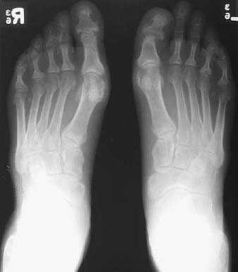
CASE 112 Peter L. Munk and Anthony G. Ryan A 41-year-old man with a skin rash presented with painful feet. Figure 112A A radiograph of the feet (Fig. 112A) shows bilateral erosion of the head of the first metatarsal, worse on the left where the head is almost completely destroyed. Corresponding but less florid erosion is present on the distal aspect of this joint, producing a characteristic deformity. Psoriatic arthropathy, demonstrating “pencil-in-cup” deformity of the left first interphalangeal joint. Psoriatic arthritis combines many features of rheumatoid arthritis, in which synovial inflammation predominates, and ankylosing spondylitis, in which ligamentous inflammation predominates. Psoriatic arthritis is of unknown etiology, although human leukocyte antigen (HLA) B27 levels are increased in 20% of patients with peripheral arthritis and 50% of patients with axial arthritis. An infectious etiology has been proposed. Psoriatic anthropathy is characterized by a profound synovitis of unknown etiology but which is associated with dysregulated angiogenesis and the presence of increased concentrations of an inflammatory serine protease. Psoriatic arthropathy most commonly affects patients between the ages of 20 and 40 years. Arthritis is said to occur in 5 to 8% of patients with psoriasis. Although there is an equal sex distribution, spine and distal interphalangeal disease is more common in men, whereas a symmetric polyarthritis is more common in women. The arthropathy precedes the characteristic rash in 10% of patients; in the majority (75%), the rash is present first. The distal interphalangeal joints of the fingers and feet are often affected; however, unlike in rheumatoid arthritis, joint involvement is frequently asymmetric. Patients report painful swollen joints. Soft-tissue swelling around the affected joints compounds the joint swelling secondary to the frequently present effusions. Joints may be stiff and immobile if bony ankylosis has occurred. Lower back pain and diminished back flexion may be present secondary to sacroiliac or spinal involvement. A spectrum of clinical severity exists. Most patients demonstrate a monoarticular pattern or an asymmetric oligoarticular pattern. Other patients have a more extensive involvement of their joints, which at times can closely mimic rheumatoid arthritis; however, rheumatoid factor is negative. A polyarthritis affects primarily the distal interphalangeal joints. A spondylarthritis closely mimics ankylosing spondylosis.
Psoriatic Arthropathy
Clinical Presentation
Radiologic Findings
Diagnosis
Differential Diagnosis
Discussion
Background
Etiology
Pathophysiology
Clinical Findings
Stages of Disease
Related posts:
Stay updated, free articles. Join our Telegram channel

Full access? Get Clinical Tree


