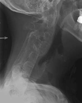
CASE 115 Anthony G. Ryan and Peter L. Munk A 33-year-old man presented with chronic back pain. Figure 115A Figure 115B Figure 115C Anteroposterior (AP) (Fig. 115A) and lateral (Fig. 115B) radiographs of the lumbar spine show bilaterally symmetric fine bridging syndesmophytes throughout the visualized spine. An AP radiograph of the pelvis (Fig. 115C) shows symmetric absence and complete bony fusion of the sacroiliac joints, with bilaterally symmetric hip joint space narrowing, axial migration of the femoral heads, erosions, and subchondral sclerosis. Ankylosing spondylitis. Other seronegative arthropathies, especially enteropathic arthropathy, are differential diagnoses. The bilateral symmetry and bridging nature of the syndesmophytes make the other seronegative spondy-loarthropathies (psoriatic arthritis and reactive arthritis) less likely. Syndesmophytosis and the degree of osseous fusion at the sacroiliac joints make rheumatoid arthritis very unlikely. The seronegative spondyloarthropathies consist of ankylosing spondylitis, psoriatic arthritis, reactive arthritis (Reiter’s disease), enteropathic spondylitis, and undifferentiated spondyloarthropathy, all of which share common clinical and radiographic features, with characteristic involvement of the sacroiliac joints, spine, and peripheral joints. The spondyloarthritides affect synovial and cartilaginous joints (including the intervertebral disks and their junctions with adjacent vertebral bodies) and entheses. Ankylosing spondylitis is seen as the prototype of the spondyloarthritides, but it may also be viewed as a potential end point of any one of the spondyloarthritides. The criteria for diagnosing one of the inflammatory spondyloarthropathies are spine pain or synovitis plus one of the following: Sacroiliitis in the presence of appropriate clinical symptoms is said to be diagnostic for ankylosing spondylitis, although the presence of typically inflammatory back pain plus at least two other typical features of spondyloarthropathy (e.g., enthesitis and uveitis) is highly predictive of early ankylosing spondylitis. The etiology of ankylosing spondylitis remains unknown; however, it is presumed to relate to an as yet unidentified chronic intracellular infectious agent, or agents, initiating a series of autoimmune phenomena in susceptible hosts. Unlike reactive arthritis (Reiter’s syndrome), no specific microbe has been identified in ankylosing spondylitis, although cross-reactivity has been demonstrated between Klebsiella and human leukocyte antigen (HLA) B27. The chief host susceptibility is related to the presence of the major histocompatibility complex HLA-B27 allele, for which over 90% of patients with ankylosing spondylosis are positive, compared with a maximum of 8% in the nonaffected population. Although the incidence of ankylosing spondylosis in the general population is ~0.01%, the incidence in patients with HLA-B27 positivity is 20%. The condition is characterized by pathological processes of bone and soft-tissue inflammation affecting bone (osteitis), synovial joints (synovitis), cartilage (chondritis), discovertebral junction (spondylodiskitis), and entheses (enthesitis), with bony repair and ossification occurring consecutively and occasionally concurrently. Two theories currently dominate the potential origin of arthritogenic peptides: an immunological theory, incriminating a cross-reaction between self-proteins (e.g., host proteoglycans) and bacterial peptides via molecular mimicry (HLA-B27 itself possibly serving as an autoantigen), and a microbiological theory, which proposes that latent bacteria residing within macrophagic or dendritic cells undergo reactivation through a process facilitated by (possibly faulty secondary to misfolding) the HLA-B27 molecule, resulting in impaired intracellular killing of bacteria. In the infectious theory, infected cells migrate from mucous membranes (e.g., inflamed intestine leading to an impaired gut-blood barrier) to the target tissues, particularly the bone marrow located near entheses, and, via HLA-mediated agents, produce inflammatory cellular and cytokine (tumor necrosis factor-α and interleukin-10) infiltrates, which in turn cause acute local inflammation. The intracellular bacteria may, at a later stage, undergo transient reactivation to cause recurrent inflammation and contribute to ongoing chronic inflammatory response. The disease typically starts between 15 and 35 years of age and affects men between three and five times as often as women. It is rare in people of African or Japanese descent but common in people with innate high levels of HLA-B27, such as the Haida Indians of British Columbia, in whom the highest prevalence of ankylosing spondylitis (4.5%) has been reported (50% of the population is HLA-B27-positive). Ankylosing spondylitis is characterized by an insidious onset of low back pain and stiffness, the latter worse in the morning. The pain is typically inflammatory in nature; that is, it disturbs sleep, is worse in the morning, and is relieved by exercise. The back pain is most commonly due to sacroiliitis, with pain in the buttocks radiating to the legs a typical feature. Occasionally, there is evidence of plantar fasciitis on presentation. Later in the course of the disease, new onset of mechanical back pain is suspicious for superimposed fracture. On examination, there is loss of lumbar lordosis and increased kyphosis, with a variable limitation of spinal flexion and reduced inspiratory chest excursion. Pelvic and chest wall tenderness is frequently demonstrable. Enthesopathy is manifest by periarticular tenderness. The manner of presentation and course tend to be different in females, who present at an older age, have a higher incidence of initial and subsequent peripheral joint disease, a higher incidence of cervical spine disease, and a milder, less progressive disease course. The appendicular involvement is more apparent clinically than radiographically. A juvenile form is also recognized in which symptoms develop before the age of 17; the most common presentation is with lower limb appendicular (asymmetric, oligoarticular), rather than axial, joint involvement. In these patients, the course tends to be progressive. The erythrocyte sedimentation rate and C-reactive protein level are typically raised on presentation in all forms. Extraskeletal manifestations include the following: These patients are more susceptible to vertebral fractures secondary to the loss of spinal flexibility and increased bone fragility. The addition of diffuse paraspinal ossification, ossified facet joints, and inflammatory osteitis creates a fused, brittle spine. Even minor trauma can produce an unstable injury as a result of disruption of the ossified supporting ligaments. Figure 115D Lateral radiograph of the cervical spine in a woman complaining of severe neck pain, having bumped her head on a sink when bending down, demonstrating a transdiscal fracture dislocation involving the posterior elements, probably representing “completion” of a stress fracture. Neurologic recovery occurred slowly after reduction and fixation. Cervical spine fractures are three times more common in patients with ankylosing spondylitis compared with the population at large. There are three recognized vertebral fracture patterns: The exact cause of the cauda equina syndrome has not yet been determined, but it may be due to demyelination, postirradiation ischemia, or compression from spinal arachnoiditis. Clinically, patients will complain of cutaneous sensory impairment of the lower limbs and perineum with sphincter disturbances. Motor impairment occurs less frequently, and associated pain is an inconstant feature. Saccular dilatation of the caudal dural sac and dorsal arachnoid diverticulae that erode the lamina and spinous processes are characteristic myelographic and CT findings. Dorsal arachnoid diverticulae occur in the lumbar spine with erosion of posterior elements. The paraspinal and psoas muscles are frequently affected secondary to gross ankylosis of the hip joints and the spine. Erosion, reactive hyperostosis, and eburnation of affected bone are identified. Fibrous or bony ankylosis of the joints is seen. The pathological hallmark of the disease is acute and chronic inflammation leading to osteitis, chondritis, synovitis, spondylodiskitis, and enthesitis, with bony repair and ossification occurring consecutively and concurrently. Endochondral ossification across the joints leads to eventual bony ankylosis. Effusions, when present, are sterile.
Ankylosing Spondylitis
Clinical Presentation
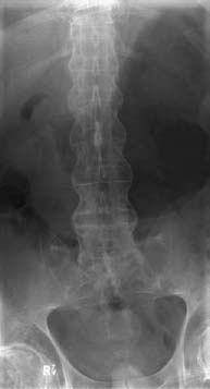
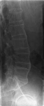
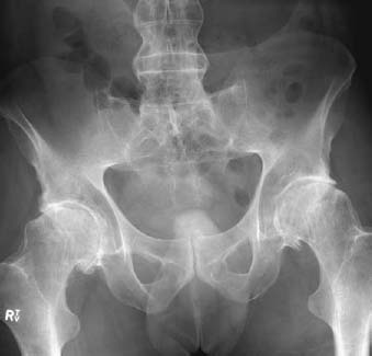
Radiologic Findings
Diagnosis
Differential Diagnosis
Discussion
Background
Etiology
Pathophysiology
Clinical Findings
Complications
VERTEBRAL FRACTURES
CAUDA EQUINA SYNDROME
MUSCULAR ATROPHY
Pathology
GROSS
MICROSCOPIC
Imaging Findings
RADIOGRAPHY
Related posts:
Stay updated, free articles. Join our Telegram channel

Full access? Get Clinical Tree


