PART VIII Other Conditions Anthony G. Ryan, Nizar Al-Nakshabandi, and Peter L. Munk A 37-year-old man presented with discomfort and swelling of the right knee that had gradually progressed over a 2- to 3-year period. This was aggravated by activity, which often produced a marked increase in swelling. Figure 119A Figures 119B Figures 119C The anteroposterior view of the knee (Fig. 119A) shows erosions involving both the femur and the tibia centered in the region of the intercondylar notch. The erosions are well defined with slightly sclerotic borders, consistent with a slowly enlarging soft-tissue mass. An MRI examination shows a large effusion distending the suprapatellar bursa (Figs. 119B, 119C). On the gradient echo image (Fig. 119B), multiple punctate foci of low signal intensity are present, consistent with areas of hemosiderin deposition. Following administration of intravenous (IV) gadolinium contrast, extensive masslike enhancement is noted (Fig. 119C). Pigmented villonodular synovitis (PVNS). Other hemosiderin-containing lesions include PVNS is a rare benign proliferative diffuse intra-articular growth of the synovium of obscure etiology, first described in 1941 and representing part of a disease spectrum that includes a localized form (giant cell tumor of the tendon sheath [GCTTS]). The more common (75 to 85%) of the two entities, GCTTS differs from PVNS in location, as it is classically extra-articular, located around the tendon sheaths of the hand, and usually measures < 2 cm in size. Although some debate persists regarding the etiology of the condition, in its diffuse form, it is widely held to be neoplastic, as evinced by the presence of mononuclear clonality and rare reported cases of metastases. Alternative etiologies suggested include an inflammatory process of unknown cause and an abnormality of local lipid metabolism. The localized form is held by some to be a local granulomatous reaction; however, a causative agent has not been identified. Repetitive trauma to the friable villi causes bleeding, resulting in synovitis, effusion, pain, and limited joint motion. Bone erosions contribute to the pain and limited range of motion.
CASE 119
Pigmented Villonodular Synovitis
Clinical Presentation
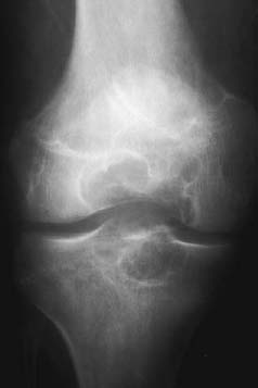
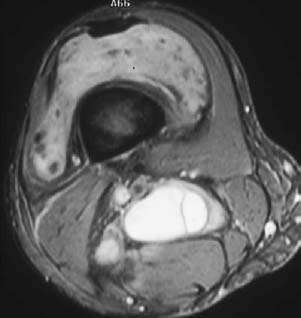
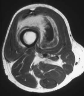
Radiologic Findings
Diagnosis
Differential Diagnosis
Discussion
Background
Etiology
Pathophysiology
Clinical Findings
Related posts:
Stay updated, free articles. Join our Telegram channel

Full access? Get Clinical Tree


