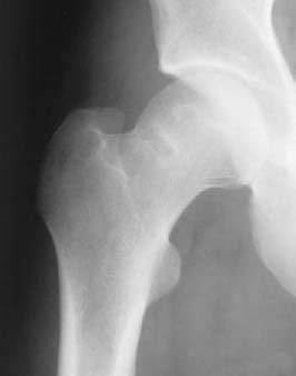
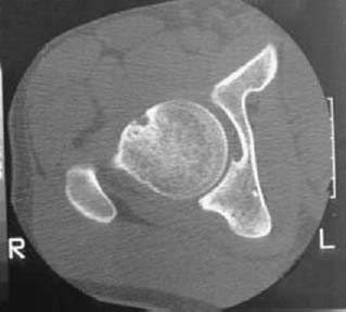
CASE 120 Anthony G. Ryan and Peter L. Munk A 26-year-old woman presented with vague hip pain. Figure 120A Figure 120B An anteroposterior radiograph of the hip (Fig. 120A) shows an apparent ill-defined lucency with a surrounding rim of sclerosis projected through the superior aspect of the femoral neck. A transaxial CT at the level of the lesion (Fig. 120B) on the radiograph confirms the presence of a lucent defect surrounded by a moderate sclerotic margin at the junction of the femoral head and neck. Synovial herniation pit (Pitt’s pit) Patients very commonly present with hip pain that often is nonspecific in character. Virtually all of these patients will go on to have radiography. A frequently encountered finding is that of a synovial herniation pit. Although unclear, mechanical stress is thought to be responsible. The level of the herniation appears to be related to the thickest portion of the capsule and the ligaments in the hip joint, where the fibers of the zona orbicularis cross and intermingle with the iliofemoral ligament. This area becomes tightest in extension and internal rotation.
Synovial Herniation Pit
Clinical Presentation
Radiologic Findings
Diagnosis
Differential Diagnosis
Discussion
Background
Etiology
Pathophysiology
Related posts:
Stay updated, free articles. Join our Telegram channel

Full access? Get Clinical Tree


