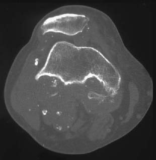
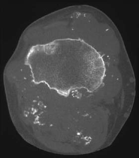
CASE 121 George Nomikos, Anthony G. Ryanm, Mark Murphey, and Peter L. Munk A 57-year-old man presented with a short history of increasing posterior right knee pain. Figure 121A A lateral radiograph of the knee (Fig. 121A) shows erosions involving the tibia and patella, as well as a soft-tissue mass in the popliteal fossa. Multiple small calcifications are also visible in the mass. Axial CT images at the level of the femoral condyle and tibial plateau (Figs. 121B, 121C) demonstrate multiple similarly sized calcified bodies in a popliteal cyst. Multiple erosions are identified in the tibia. Primary (idiopathic) synovial chondromatosis. Figure 121B Figure 121C Primary synovial chondromatosis (PSC) is a rare condition characterized by formation of multiple cartilaginous or osteocartilaginous bodies, usually of similar size, in a synovial joint. It must be distinguished from secondary (osteo)chondromatosis, in which multiple intra-articular bodies (IABs) are a result of another primary joint abnormality, such as osteoarthritis, osteonecrosis, neuropathic disease, trauma, osteochondritis dissecans, or rheumatoid arthritis. The IABs may be mineralized or nonmineralized. Synovial osteochondromatosis is a condition of unknown etiology in which synovial tissue undergoes benign reactive cartilaginous metaplasia to form multiple (osteo)cartilaginous nodules ranging from 1 mm to 3 cm in size. These osteochondral fragments in primary synovial chondromatosis tend to be similar in size and innumerable, unlike secondary osteochondromatosis, in which the fragments are usually of varying size and few in number. These chondral fragments may be free in the joint or may become reattached to synovium.
Synovial Osteochondromatosis
Clinical Presentation

Radiologic Findings
Diagnosis
Differential Diagnosis
Discussion
Background
Etiology
Clinical Findings
Related posts:
Stay updated, free articles. Join our Telegram channel

Full access? Get Clinical Tree


