CASE 125 Peter L. Munk, Anthony G. Ryan A 37-year-old man presented with a complaint of lower back pain. Figure 125A Figure 125B An anteroposterior radiograph of the pelvis (Fig. 125A) shows multiple surgical clips in the left lower quadrant, marked osteoporosis of both femoral heads, and apparent healing fractures of the pubic rami bilaterally. A coned view of the distal hands (Fig. 125B) shows osteoporosis with cortical irregularity and subperiosteal lucency on the radial aspects of the phalanges and subtle lucency of the distal phalangeal tufts. Renal osteodystrophy (secondary hyperparathyroidism). The pubic abnormalities are Looser’s zones: pseudofractures secondary to osteomalacia. The surgical clips are in relation to a renal transplant (failed). None. Renal osteodystrophy is a constellation of clinical, pathologic, and radiologic manifestations seen in patients with chronic renal disease. The radiological manifestations are diverse and fall into several categories. With the increasing utilization of hemodialysis and aggressive management of patients with renal disease, patients with imaging manifestations of renal osteodystrophy are increasingly encountered. These changes are in many cases reversible following renal transplantation, although they may take years to regress. These changes can be broadly categorized under several headings. The osseous changes due to renal osteodystrophy are for the most part due to secondary hyperparathyroidism. Patients with renal failure often undergo hypertrophy of the hyperparathyroid glands induced by hyperphosphatasemia due to decreased renal excretion. Increased circulating levels of parathyroid hormone (PTH) then alters bone metabolism, leading to a protean constellation of changes. In particular, PTH promotes the development of osteoclasts, osteoblasts, and osteocytes, giving rise to bone resorption, periosteal reactions, and brown tumors. Osteosclerosis, osteoporosis, and osteomalacia may all occur; hence the sobriquet “active bones disease.” Osteoclastic bone resorption is probably the most consistent feature and may affect any portion of the bone, that is, subperiosteal, cortical, subchondral, trabecular, endosteal, or at entheses. Patients with renal failure due to chronic disease affecting the glomeruli also develop alterations in vitamin D metabolism, frequently showing decreased sensitivity to the vitamin. This in turn also alters bone metabolism, with osteomalacia or rickets developing. Metabolic interactions become very complex, and often several manifestations of disease are present simultaneously.
Hyperparathyroidism / Renal Osteodystrophy
Clinical Presentation
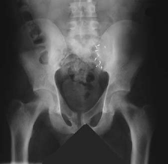
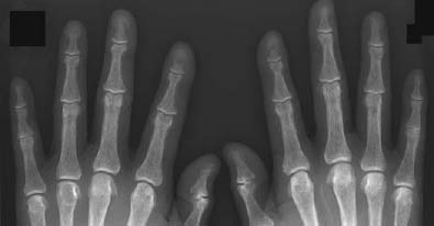
Radiologic Findings
Diagnosis
Differential Diagnosis
Discussion
Background
Etiology
Pathophysiology
Clinical Findings
Complications
Pathology
GROSS
Related posts:
Stay updated, free articles. Join our Telegram channel

Full access? Get Clinical Tree


