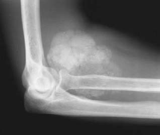
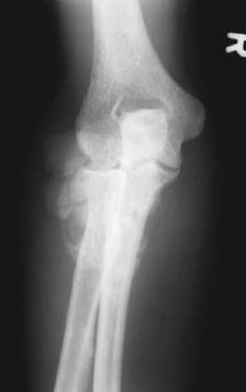
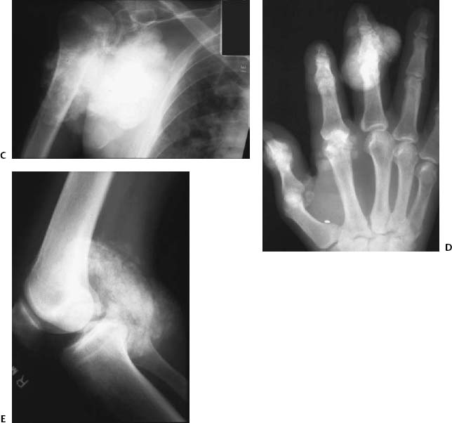
CASE 126 Brian Edward Reeves, Anthony G. Ryan, Peter L. Munk, and Thomas Pope A 26-year-old woman presented with painless stiffness and swelling in her right elbow. Figure 126A Figure 126B Lateral (126A) and anteroposterior (126B) radiographs of the right elbow show a lobulated homogeneous, calcific mass within the antecubital fossa. The articular space is normal, and there is no erosion of bone. Tumoral calcinosis. Idiopathic tumoral calcinosis is a rare and poorly understood entity consisting of calcium salt deposition in the extracapsular soft tissues adjacent to joints. It occurs most commonly in the first or second decade, predominantly affects African Americans, and is seen more frequently in men. One third of the cases are thought to be familial, and the inheritance is likely autosomal dominant with variable expression. There is some evidence suggesting that tumoral calcinosis may be caused by an inborn abnormality of phosphorus metabolism; other metabolic defects, collagen vascular disorders, and trauma have also been suggested as pathogenic mechanisms. Figures 126C–126E
Tumoral Calcinosis
Clinical Presentation
Radiologic Findings
Diagnosis
Differential Diagnosis
Discussion
Background
Etiology
![]()
Stay updated, free articles. Join our Telegram channel

Full access? Get Clinical Tree


