CASE 128 Anthony G. Ryan and Peter L. Munk A 60-year-old man presented with bilateral painful hands. Figure 128A An anteroposterior radiograph of both hands (Fig. 128A) shows joint space reduction affecting the metacarpophalangeal, radiocarpal, and interphalangeal joints; diffuse osteopenia; and prominent hooked osteophytes on the radial aspect of the second and third metacarpals, the heads of which are enlarged by the presence of the osteophytes. Variable lucencies, marginated by thin, sharply defined rims of sclerosis, are evident within the metacarpal heads. Chondrocalcinosis is evident in the triangular fibrocartilage bilaterally. The scapholunate interval is of normal size. Hemochromatosis arthropathy. Hemochromatosis may occur as a primary or secondary (acquired) condition. The primary, idiopathic form is transmitted as an autosomal recessive trait, with only homozygotes demonstrating the clinical condition, or via indeterminate inheritance (e.g., there is an abnormal iron-loading gene present in thalassemia and sideroblastic anemia, resulting in increased iron absorption with a normal dietary load). Acquired hemochromatosis occurs secondary to Both primary and secondary causes result in deposition of excessive iron in the liver, pancreas, spleen, gastrointestinal tract, kidney, gonads, heart, and endocrine glands, resulting in fibrosis, which leads to eventual organ failure. Iron deposition in the synovium leads to an arthropathy in 30 to 50% of cases. Synovitis is also caused and/or exacerbated by secondary calcium pyrophosphate deposition (CPPD), leading to chondrocalcinosis, most frequently found in the triangular fibrocartilage of the wrist, the menisci, the anulus fibrosus, the ligamentum flavum, the symphysis pubis, the Achilles’ tendon, and the plantar fascia. The calcification is due to deposition of calcium pyrophosphate crystals, perhaps resulting from iron inhibition of pyrophosphatase.
Hemochromatosis
Clinical Presentation
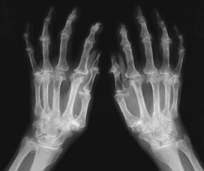
Radiologic Findings
Diagnosis
Differential Diagnosis
Discussion
Etiology
Pathophysiology
Clinical Findings
Related posts:
Stay updated, free articles. Join our Telegram channel

Full access? Get Clinical Tree


