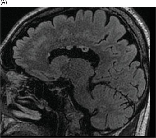
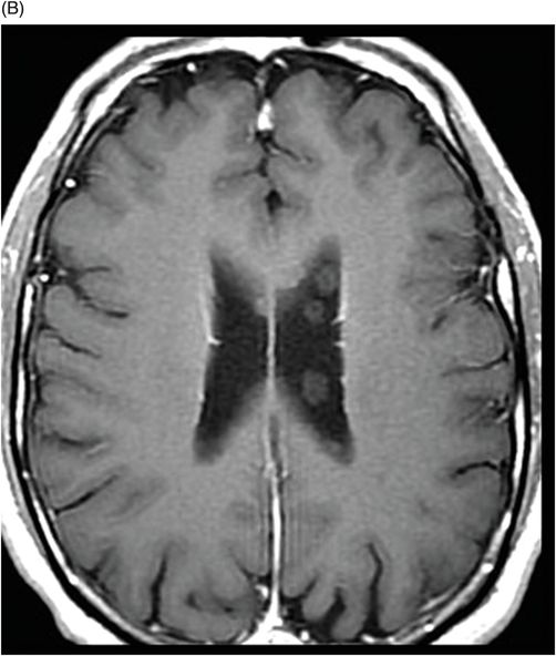
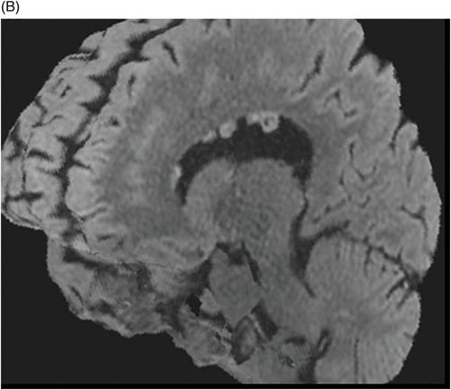
(A) Sagittal volumetric FLAIR and (B) Contrast-enhanced T1WI through the level of the lateral ventricles.
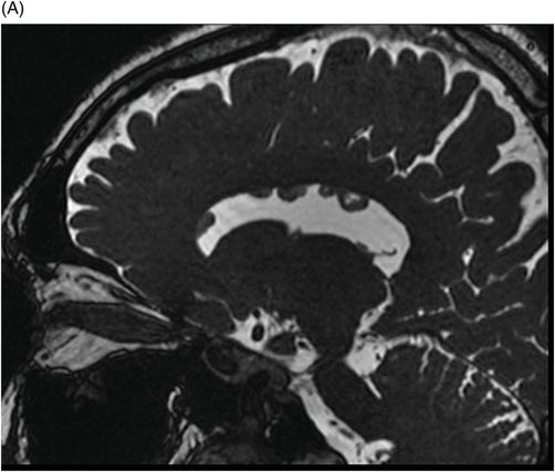
(A) Axial and (B) Sagittal 3D-volumetric, FLAIR reconstructions through the level of the lateral ventricles.
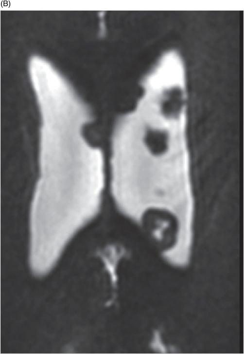
(A) Sagittal and (B) Axial 3D-CISS-FIESTA MR images through the level of the lateral ventricles.
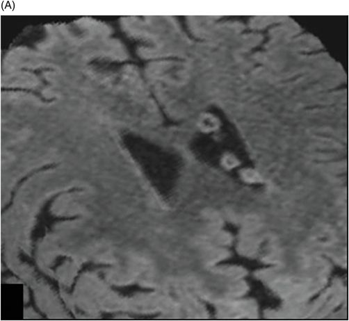
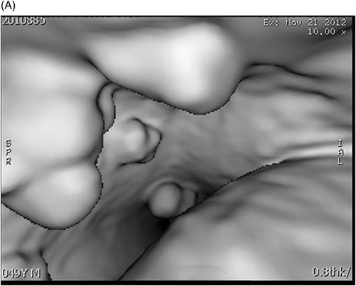
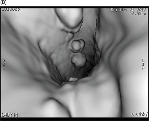
(A–B) Intraventricular, virtual endoscopic-3D reconstructed MR images through the level of the left lateral ventricle.
Ring-Shaped Lateral Ventricular Nodules
Primary Diagnosis
Ring-shaped lateral ventricular nodules
Differential Diagnoses
Subependymal heterotopia
Subependymal nodules in tuberous sclerosis complex
Subependymoma
Imaging Findings
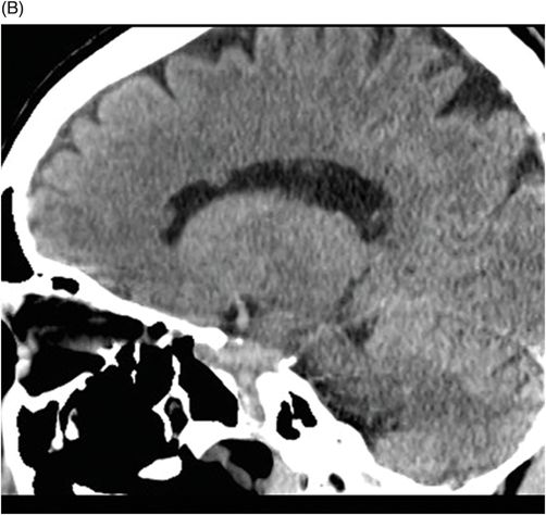
Fig. 130.1: (A) Axial and (B) Sagittal unenhanced CT images through the level of the lateral ventricles showed small, nodular lesions in the left lateral ventricle, isodense relative to gray matter. One of the lesions demonstrated a central portion that was isodense to CSF. Fig. 130.2: (A) Sagittal volumetric FLAIR MR image through the level of the lateral ventricles showed slight hypersignal, relative to the parenchyma of the lesions’ ring portion. An isointense signal of the lesions’ core portion, relative to CSF, was noted. (B) T1W contrast-enhanced image did not show contrast enhancement of the lesions. Fig. 130.3: (A) Axial and (B) Sagittal 3D-volumetric FLAIR reconstructions better demonstrated the hypersignal of the lesions’ ring portion and the isointense signal of their core portion, relative to CSF. Fig. 130.4: (A) Sagittal and (B) Axial 3D-CISS-FIESTA MR images showed ring-shaped, nodular lesions in both lateral ventricles and their core portion was isointense relative to CSF. Fig. 130.5: (A) and (B) Intraventricular, virtual endoscopic-3D reconstructed MR images through the level of the left lateral ventricle showed the nodular lesions and their round, oval, lobulated configurations and their preferential location in the roof of the body of the lateral ventricle.


Stay updated, free articles. Join our Telegram channel

Full access? Get Clinical Tree


