Fig. 20.1
Pneumococcal meningoencephalitis in a 57-year-old patient. (a) Axial FLAIR and (b) axial T2 TSE images show hyperintensity of right frontoparietal gray matter and leptomeningeal spaces. (c) b1000 DWI and (d) ADC map show restricted diffusivity of leptomeninges and subarachnoid space, as compared to the contralateral side. (e, f) axial T1-weighted TSE images, respectively, without and with fat suppression technique, before and after contrast agent administration, show diffuse right leptomeningeal and cortical frontoparietal enhancement
MRI also plays an important role in the diagnosis and monitoring of meningitis complications such as hydrocephalus, cerebritis, abscess, cranial nerves inflammation, venous thrombosis, vasculitis, infarct, ventriculitis, and subdural empyema [8].
In a recent study [9], differences of DTI indices in several limbic regions and in the white matter close to the globus pallidus were found in patients with chronic neuropsychological sequelae of chronic meningitis with respect to healthy subjects. Authors argued that their results suggest that white matter alterations may be involved in the psychopathology and pathophysiology of chronic meningitis. To our knowledge, there are no other studies investigating the utility of advanced MRI techniques in the diagnosis and monitoring of meningitis.
20.3 Encephalitis
Encephalitis is an acute inflammation of the brain. The etiology is most frequently viral. Childhood and adult herpes simplex virus (HSV) encephalitis is usually due to HSV-1 (90 %). Reactivation of dormant HSV-1 in the trigeminal ganglion precipitated by various factors (trauma, immunosuppression, hormonal fluctuations, stress) causes necrotizing and hemorrhagic involvement of white and gray matter that primarily affect the limbic system. Frontal, parietal, and occipital cortex can also be involved, while basal ganglia are usually spared. Lesions are usually asymmetric and bilateral.
Conventional MRI signs include gray and subcortical white matter hyperintensity on T2-weighted sequence with corresponding signal decrease on T1-weighted sequence, loss of gray-white matter junction, sulci effacement, and focal hypointensities on T2* images. Enhancement is best seen 1 week after initial symptoms; it may be gyral, leptomeningeal, ring, or diffuse.
Restricted diffusivity of the affected cortex is a more sensitive sign of HSV encephalitis with respect to other conventional imaging signs. However, 14 days after symptom onset, diffusivity in the cerebral cortex is no longer restricted, while T2WI and FLAIR abnormalities persist (Fig. 20.2).

Fig. 20.2
HSV encephalitis in a 64-year-old patient. (a) Axial FLAIR and (b) coronal T2 TSE image show left temporal lobe gray and subcortical white matter hyperintensity, with brain swelling and sulci effacement. (c) b1000 DWI showing restricted diffusivity of left temporal cortex. (d, e) axial T1-weighted TSE images before and after contrast agent administration show cortical enhancement
20.4 Cerebritis, Abscesses, and Granulomas
Infective focal brain lesions include abscesses and granulomas. A pyogenic infection of the brain can cause the formation of an abscess, through the activation of a purulent inflammatory process evolving through four stages: early cerebritis, late cerebritis, early capsule formation, and late capsule formation. Contrarily, chronic granulomatous inflammation mediated by cells of the mononuclear phagocyte system leads to the formation of well-demarcated focal lesions, containing cellular debris, macrophages, lymphocytes, plasma cells, and fibroblasts. Parasitic and fungal granulomas contain also eosinophils.
20.4.1 Bacterial
Most frequent pathogens responsible of abscesses formation are Streptococcus spp., Enterobacteriaceae, anaerobes, and staphylococci. Brain involvement can be direct from a contiguous site of infection (otitis media, mastoiditis, paranasal sinusitis, dental infection, head trauma, neurosurgical procedure) or indirect by hematogenous dissemination from distant sites of infection.
In the early cerebritis stage (days 1–3), a central core of coagulative necrosis is surrounded by a perivascular infiltration of inflammatory cells and marked edema; conventional MRI shows an ill-defined mass, hyperintense on T2-weighted sequence, and isointense-hypointense on T1-weighted sequence with a patchy enhancement after contrast agent administration.
In the late cerebritis stage (days 4–9), the necrotic purulent center and the surrounding inflammatory infiltrate of macrophages and fibroblasts are divided by a thin capsule of fibroblasts; conventional MRI shows a central core hypointense/hyperintense on T1-/T2-weighted sequences, a peripheral rim mildly hyperintense/hypointense on T1-/T2-weighted sequences, and a marked but irregular rim enhancement after contrast agent administration.
In the early capsule formation stage (days 10–13), a well-demarcated capsule is surrounded by marked edema; conventional MRI shows a ring-enhancing lesion with a typical smooth inner margin of the ring, a hypointense signal of the rim on T2-weighted images, and a markedly restricted diffusivity of the central core (Fig. 20.3).

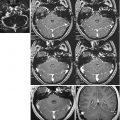
Fig. 20.3
Pyogenic abscess in a 71-year-old patient. (a) Axial FLAIR and (b) axial T2 TSE images show a right frontal expanding lesion surrounded by large edema. Note the hypointense ring. (c) b1000 DWI and (d) ADC map show a markedly restricted diffusivity of the internal cavity. (e, f) Axial T1-weighted TSE images before and after contrast agent administration show smooth margins of the inner and outer layers of the enhanced ring
In the late capsule formation stage (more than 14 days), a well-formed necrotic core and surrounding marked gliosis with large numbers of reactive astrocytes are divided by a dense collagenous capsule; conventional MRI shows thickening of the enhancing rim and reduction of edema signs with respect to the previous stage.
Restricted diffusivity in the central core of a ring-enhancing lesion is by far the most specific advanced MRI technique sign for pyogenic brain abscess [10] opposite to non-pyogenic lesions that show hypointense or mixed signal on b1000 DWI images. However, as exceptions to the rule, histologically proven pyogenic brain abscesses that didn’t show restricted diffusivity of their central core were reported [11, 12]. Furthermore, metastases and glioblastomas with ring enhancement and restricted diffusivity of their central core were also reported [11].
In unclear cases, additional information can be gathered from other advanced MRI techniques (Fig. 20.4).

Fig. 20.4
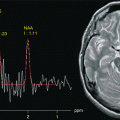
Same case as in Fig. 20.3. (a) 3 T single-voxel long-echo time (TE = 144 ms) water–suppressed 1H-MRS spectrum shows lactate and lipid peaks not only in the internal cavity but also in the perilesional edema. (b) 3 T dynamic susceptibility contrast-enhanced cerebral blood volume color map shows low CBV values in the ring and in the perilesional edema. (c) 3 T DTI color map shows that frontal forceps fibers are not interrupted
Fractional anisotropy (FA) in the central cavity of a ring-enhancing lesion is higher in pyogenic abscesses with respect to glioblastomas and metastases [13–14]. MRS reveals amino acids only in the central cavity of pyogenic abscesses and not in the central cavity of necrotic tumors [15]. Lactate cytosolic amino acids with/without succinate, acetate, alanine, and glycine peaks are markers of abscess [15]. Cerebral blood volume (CBV), if measured in the areas of ring enhancement and perilesional edema, is generally higher in malignant lesions than in pyogenic abscesses [16, 17].
Related posts:
Stay updated, free articles. Join our Telegram channel

Full access? Get Clinical Tree


