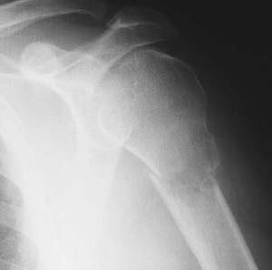
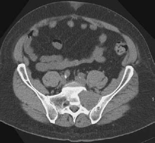
CASE 46 George Nomikos, Anthony G. Ryan, Peter L. Munk, and Mark Murphey A 55-year-old patient presented with left shoulder pain. Figure 46A Figure 46B A single view of the shoulder (Fig. 46A) shows a pathologic fracture through a lytic destructive lesion of the left proximal humerus. The lesion involves the epiphysis, metaphysis, and diaphysis of the left proximal humerus. The lesion shows a relatively wide zone of transition. There is extensive bone destruction. No mineralized matrix is identified in the lesion. A single image from a CT scan through the pelvis in the same patient is shown in Fig. 46B, demonstrating a soft-tissue mass within the left sacral ala with extensive medullary replacement and cortical breakthrough anteriorly. Metastatic renal cell carcinoma. After the lung and liver, bone is the third most common site of metastatic disease. Approximately 30% of patients with a primary carcinoma develop bone metastases, and ~70% of these patients suffer from pain related to the osseous disease. The most common malignancies to give rise to osseous metastases are in the kidney, prostate, breast, lung, and thyroid. Most osseous metastases occur in the axial skeleton (80%), with the spine representing the single most common site. This is likely related to the large amount of hematopoietic marrow in this portion of the skeleton. Malignancies may spread to bone via several different routes, including direct extension, lymphatic spread, hematogenous dissemination, and intraspinal spread via the cerebrospinal fluid. Although these lesions may be asymptomatic and only discovered incidentally, pain at the site of metastasis (which may be due to pathologic fracture) and neurologic compromise secondary to spinal involvement are the most common presentations. Histology reflects the primary malignancy. Skeletal metastases may be osteolytic, osteosclerotic, or mixed. Tables 46-1 to 46-3 summarize the most common osteolytic (Table 46-1), mixed (Table 46-2), and osteosclerotic (Table 46-3) metastatic lesions. Lesions are often multiple; however, solitary osseous metastases may occur. Periosteal reaction is less commonly seen in the setting of metastatic disease to bone than in the case of primary bone tumors. Certain tumors have a predilection to cause prominent osseous expansion, particularly renal, thyroid, and liver carcinomas. Large osteoblastic lesions may be identified in patients with metastatic prostate cancer.
Carcinoma
Clinical Presentation
Radiologic Findings
Diagnosis
Differential Diagnosis
Discussion
Background
Etiology
Pathophysiology
Clinical Findings
Complications
Pathology
Imaging Findings
RADIOGRAPHY
| Thyroid cancer |
| Renal cancer |
| Adrenal cancer |
| Uterine cancer |
| Gastrointestinal cancers |
| Melanoma |
| Hepatoma |
| Squamous cell carcinoma of the skin |
| Certain head and neck cancers |


