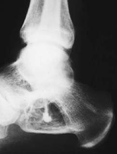
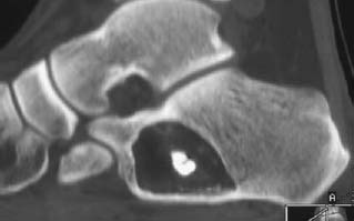
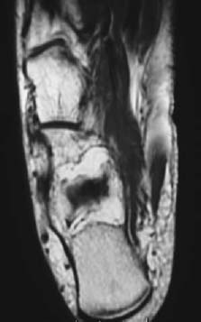
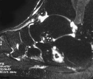
CASE 53 George Nomikos, Mark Murphey, Anthony G. Ryan, and Peter L. Munk A 32-year-old man presented with foot pain. Figure 53A Figure 53B Figure 53C Figure 53D The lateral radiograph of the ankle (Fig. 53A) shows a geographic lytic lesion in the anterior calcaneus with a narrow zone of transition and significant surrounding sclerosis. There is a central area of dense mineralization in the lesion. There is no evidence of pathologic fracture or aggressive bone destruction. CT (Fig. 53B) shows a well-marginated lesion arising in the anterior calcaneus. Note the sclerotic margin and central dense focus of mineralization. The predominant internal attenuation of the lesion is identical to subcutaneous fat. Axial T1- and sagittal fat-suppressed T2-weighted images demonstrate that the majority of the lesion follows fat signal intensity on the T1- and T2-weighted images (Figs. 53C, 53D); however, there is central fluid signal intensity in the lesion, around the area of mineralization. Involuting intraosseous lipoma. Intraosseous lipomas are uncommon benign bone tumors, accounting for ~0.1% of bone tumors. However, in our opinion and experience, they are much more frequent. Although the radiographic appearance of lesions in the calcaneus is often diagnostic, lesions in other locations may have a more variable appearance (see differential diagnosis list above). Three stages have been described: lesions composed virtually entirely of mature fat cells; lesions composed of mature fat cells, fat necrosis, and mineralization; and lesions demonstrating cyst formation, myxoid degeneration, and reactive woven bone production.
Intraosseus Lipoma
Clinical Presentation
Radiologic Findings
Diagnosis
Differential Diagnosis
Discussion
Background
Clinical Findings
Related posts:
Stay updated, free articles. Join our Telegram channel

Full access? Get Clinical Tree


