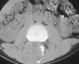
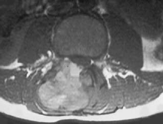
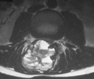
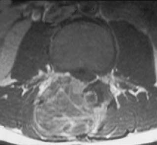
CASE 56 George Nomikos, Anthony G. Ryan, Peter L. Munk, and Mark Murphey A 13 year-old-boy presented with a history of 2 months of back pain. He reported that the pain radiated down his right lateral thigh and worsened with activity. Figure 56A Figure 56B Figure 56C Figure 56D The CT scan (Fig. 56A) shows a markedly expansile lesion arising in the posterior elements of the spine at the L2 level. Multiple fluid levels are identified in the lesion on CT and no mineralized matrix is seen. The lesion causes mild compression of the thecal sac. The axial T1- and T2-weighted images (Figs. 56B, 56C) demonstrate multiple fluid levels in all areas of the lesion with no identifiable solid component. Despite its marked expansion, the mass appears well circumscribed. The postcontrast MRI (Fig. 56D) shows peripheral and septal enhancement of the lesion. No solid enhancing nodular component is identified. Primary aneurysmal bone cyst (ABC). ABCs are reactive, not neoplastic, lesions composed of multiple blood-filled spaces. Primary ABCs represent lesions not associated with an underlying tumor, and secondary ABCs represent lesions associated with an antecedent tumor (Table 56–1). ABCs are considered secondary in from 1 to 30% of cases. An ABC is thought to arise when there is disruption in the normal vascular system of the host bone, which often occurs secondary to trauma or tumor. There is often a history of trauma to the affected area in cases of primary ABC, particularly in subperiosteal or intracortical lesions. This alteration in the normal hemodynamics leads to rapid enlargement of multiple blood-filled vascular spaces as well as proliferation of the osseous mesenchymal tissue. The patient in Fig. 56E presented initially with uncomplicated fractures of the radius and ulna. Six years later, the patient presented with swelling in the left arm. The radiograph at that time showed a markedly expansile lytic lesion arising in the radius at the site of the prior fracture (Fig. 56F), representing a primary ABC. The multiple fluid levels within the ABC are well demonstrated on the MRI (Fig. 56G).
Aneurysmal Bone Cyst
Clinical Presentation
Radiologic Findings
Diagnosis
Differential Diagnosis
Discussion
Background


