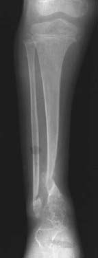
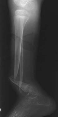
CASE 59 George Nomikos, Anthony G. Ryan, Peter L. Munk, and Mark Murphey A 15-year-old boy presented with deformity of the right, slowly progressive over a period of several years. Figure 59A Figure 59B Anteroposterior (Fig. 59A) and lateral (Fig. 59B) radiographs of the lower leg in this skeletally immature patient demonstrate chronic pseudarthroses of the distal tibial and fibular shafts. The visualized bones are gracile and osteopenic. Neurofibromatosis Type 1 (von Recklinghausen’s disease). Congenital or infantile pseudarthrosis of the tibia may be seen sporadically in the general population and may also been seen in association with osteofibrous dysplasia. However, ~50% of all cases of congenital or infantile pseudarthrosis occur in the setting of neurofibromatosis type 1 (NF1). NF1 is a common genetic disease, with a frequency of 1 case in every 2500 to 3000 births, and is the most common of the phakomatoses. It is inherited in an autosomal dominant pattern; however, up to 50% of new cases are thought to arise from spontaneous mutations. It is a mesodermal dysplasia that affects multiple organ systems. Advanced paternal age is thought to be one potential predisposing factor. An abnormality in the pericentromeric region of chromosome 17, which is responsible for coding for the protein neurofibromin, is the underlying genetic abnormality.
Neurofibroma
Clinical Presentation
Radiologic Findings
Diagnosis
Differential Diagnosis
Discussion
Background
| Six or more café-au-lait spots > 5 mm in diameter in children or > 15 mm in diameter in adults |
| Two or more neurofibromas or one plexiform neurofibroma |
| Optic glioma |
| Two or more iris hamartomas (Lisch nodules) |
| Axillary or inguinal freckling |
| Distinctive osseous lesions (sphenoid dysplasia, pseudarthrosis, vertebral scalloping, dystrophic scoliosis) |
| A first-degree relative with neurofibromatosis type 1 as diagnosed by these criteria |
Clinical Findings
The clinical criteria for establishing a diagnosis of NF1 are summarized in Table 59–1. The diagnosis can be established when two or more of these criteria are present. The classic triad of NF1 consists of cutaneous lesions, skeletal deformity, and mental deficiency. Café-au-lait spots not only are seen in patients with NF1 but also may be seen in the setting of fibrous dysplasia and tuberous sclerosis.
The orthopedic abnormalities seen with NF1 are the following:
Related posts:
Stay updated, free articles. Join our Telegram channel

Full access? Get Clinical Tree


