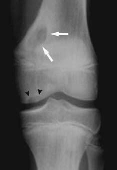
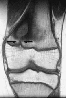
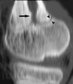
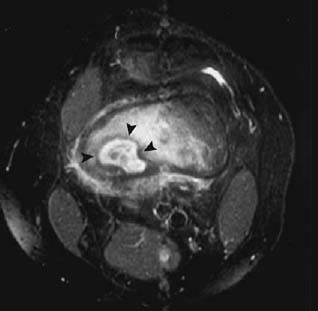
CASE 69 Sam Y. Chun, Ali Islam, Alison Spouge, Anthony G. Ryan, and Peter L. Munk A 15-year-old boy with knee pain diagnosed with osteochondritis dissecans (OCD) of the knee was sent for MRI evaluation of the OCD. Figure 69A Figure 69B Figure 69C Figure 69D An anteroposterior radiograph of the knee (Fig. 69A) shows an area of OCD in the midmedial femoral condyle (arrowheads). A large geographic lytic area in the posteromedial distal femoral metaphysis is also evident (arrows), with marginal sclerosis fading peripherally. A coronal T1 spin-echo image (Fig. 69B) shows a corresponding area of extensive low signal intensity with a small track (arrows) extending caudally to the physeal plate. A sagittal computed tomography reformat (Fig. 69C) shows the lower end of the low density lesion (arrow) to be in continuity with the physis, which is open posteriorly (arrowheads). Axial inversion recovery (Fig. 69D) shows the lesion to have a heterogeneous appearance (arrowheads) with areas of central intermediate and high signal intensity, surrounded by a low signal intensity rim with surrounding bone marrow and soft-tissue edema. Brodie’s abscess.
Brodie’s Abscess (Subacute Osteomyelitis)
Clinical Presentation
Radiologic Findings
Diagnosis
Differential Diagnosis
Discussion
Background
Related posts:
Stay updated, free articles. Join our Telegram channel

Full access? Get Clinical Tree


