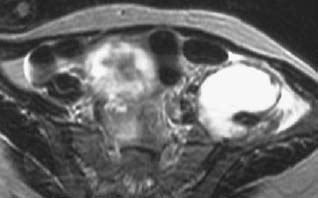
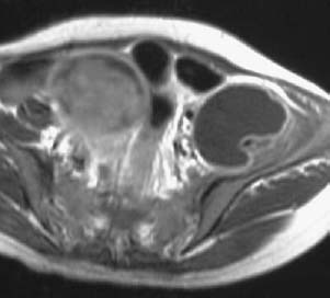
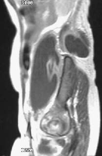
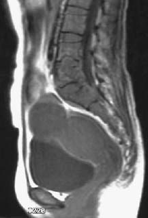
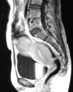
CASE 73 Ali Islam, Alison Spouge, Anthony G. Ryan, and Peter L. Munk A 38-year-old woman presented with left lower quadrant abdominal pain and a background of chronic back pain. Clinical examination revealed a palpable mass within the left lower quadrant and a vague swelling in her back just cephalad to her iliac crest. Figure 73A Figure 73B Figure 73C Figure 73D Figure 73E A transaxial T2-weighted image (Fig. 73A) through the pelvis shows a high signal collection, which on T1-weighted image postgadolinium (Fig. 73B) reveals a hypointense center and an enhancing rim. A parasagittal T1-weighted image postgadolinium (Fig. 73C) shows the same collection in longitudinal section. Extension of the collection posterior to the spine above the iliac crest can be seen. A sagittal T1-weighted image (Fig. 73D) reveals an ill-defined lobular lesion within the vertebral body of L5, which crosses the disk space superiorly into L4 and inferiorly into the back of the S1 vertebral body. Although the center of the lesion is hypointense, it demonstrates some internal heterogeneity. Most of the L4/L5 disk space is obliterated. A sagittal T1-weighted image postgadolinium (Fig. 73E) shows rim enhancement of the lesion with heterogeneous enhancement of the surrounding vertebral bodies. Pott’s disease (tuberculosis [TB] of the spine). Tuberculosis infects nearly 2 billion people worldwide, of which musculoskeletal infection accounts for nearly 2%. Tuberculosis of the spine (accounting for up to 60% of all musculoskeletal tuberculosis), or Pott’s disease, as described by Percival Pott in 1779, is one of the most dangerous forms of tuberculous infection, as it may cause paraplegia and vertebral body destruction and collapse. It accounts for roughly half of the cases of musculoskeletal infection. Tuberculosis is endemic in the population of many developing nations, and Pott’s disease is common in children living in these areas. There is no gender predilection. In the developed world, risk factors for Pott’s disease include human immunodeficiency virus (HIV), immunosuppression, alcoholism, homelessness, drug abuse, and residence in long-term care facilities and prisons. Pott’s disease is characterized by infection with the acid-fast bacillus Mycobacterium tuberculosis
Pott’s Disease
Clinical Presentation
Radiologic Findings
Diagnosis
Differential Diagnosis
Discussion
Background
Etiology
![]()
Stay updated, free articles. Join our Telegram channel

Full access? Get Clinical Tree


