PART V Avascular Bone
CASE 76
Avascular Necrosis of the Femoral Head
Anthony G. Ryan Peter L. Munk
Clinical Presentation
A 45-year-old man presented with chronic left hip pain exacerbated by exercise, status post subcapital fracture, which had been acutely pinned and appeared radiographically healed. The patient’s range of motion was limited by pain.
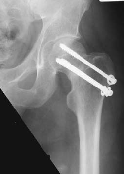
Figure 76A
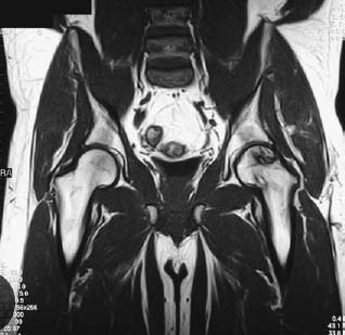
Figure 76B
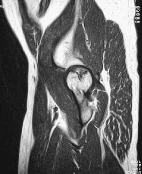
Figure 76C
Radiologic Findings
An anteroposterior radiograph of the left hip with fixation screws in situ shows some joint space narrowing and acetabular articular sclerosis, but no other significant abnormality (Fig. 76A). A coronal T1-weighted image taken 3 months later (Fig. 76B) shows a crescentic low signal intensity line undermining the superolateral femoral head with heterogeneity of the cephalad fragment, which itself contains multiple low signal intensity lines. Loss of continuity of the cortical margin is evident laterally. Irregularity of the articular surface of the femoral head and adjacent acetabular articular surfaces is evident.
Similar findings are demonstrated on the accompanying sagittal T1-weighted image (Fig. 76C), which shows the loss of joint space and articular surface irregularity with greater conspicuity. The prominent, well-demarcated low signal intensity line assumes a triangular margin in this plane.
Diagnosis
Avascular necrosis (AVN) of the femoral head.
Differential Diagnosis
None.
Discussion
Etiology
Interruption of the blood supply to bone may occur secondary to trauma to, or obstruction of, the vessels supplying the bone by intravascular elements, vasculitis, or extravascular compromise.
In the adult, traumatic arterial interruption is typically caused by fracture of the femoral neck or, less commonly, by hip joint dislocation. Intravascular causes of arterial obstruction include hemoglobinopathies and caisson disease (“the bends”).
Vasculitic causes include connective tissue disorders and radiation exposure. Increased intraosseous pressure may arise secondary to accumulation of abnormal cells, such as histiocytes in Gaucher’s disease on fat cells secondary to steroid usage.
The mechanism by which alcohol abuse causes AVN has not been clearly identified. It is thought that pancreatitis causes AVN by a combination of the above, for example, vasculitis associated with abnormal intravascular cells.
A cause for avascular necrosis is not identified in as many as 25% of cases.
The femoral head is at particular risk for necrosis by virtue of the tenuous blood supply to the femoral epiphysis via the medial circumflex and lateral epiphyseal arteries.
Pathophysiology
The hallmark of AVN is, as its name suggests, an ischemic insult producing interruption (of varying duration, depending on the etiology) of the blood supply to the affected bone or portionof bone. The radiologic features of fragmentation, collapse, sclerosis, and eventual reossification of the involved bone correlate well with the pathologic progress and repair of the resultant osteonecrosis. Disruption of the blood supply, sufficient to cause anoxia, causes death of hematopoietic cells in 6 to 12 hours, bone cell death within 12 to 48 hours, and marrow fat cell death after 48 to 60 hours.
Related posts:
Stay updated, free articles. Join our Telegram channel

Full access? Get Clinical Tree


