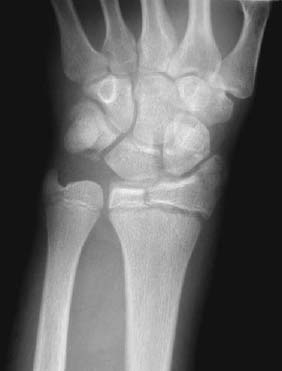
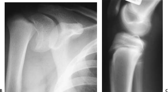
PART VI Trauma Hema N. Choudur, Anthony G. Ryan, Peter L. Munk, and Laurel O. Marchinkow A young boy presented with severe wrist pain after having fallen on his outstretched hand while playing soccer. Figure 79A An anteroposterior radiograph of the wrist (Fig. 79A) shows a fracture of the distal radial epiphysis with widening of the radial aspect of the distal radial growth plate. A fracture of the triquetrum is also evident. Salter-Harris fracture (type 3). None. Salter-Harris fractures are fractures of the growth plate and are therefore seen only in the pediatric population, prior to growth plate closure. They are classified as fractures of the physis with variable involvement of the epiphysis and metaphysis. These fractures usually occur secondary to horseplay or sports injuries. Nutrition is supplied to the growth plate from the distal to the proximal epiphyseal end by the epiphyseal vessels. The growth plate itself is avascular. The cartilage cells transform as they progress from the germinal layer toward the metaphysis to form the zone of provisional calcification. Neovascularization occurs from the metaphyseal end of the growth plate. Fractures that result in apposition and consequently compression of the vascular beds on both sides of the growth plate halt further growth. Those that occur through the growth plate with no fracture displacement do not impede growth. Disruption of the vascularity at either end of the growth plate may hamper growth. The child usually complains of pain or points to the affected bone following the injury. There is swelling and variable bruising at the site of the injury.
CASE 79
Salter-Harris Fractures
Clinical Presentation

Radiologic Findings
Diagnosis
Differential Diagnosis
Discussion
Etiology
Pathophysiology
Clinical Findings
Complications
Radiology Key
Fastest Radiology Insight Engine


