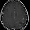

(A–B) Axial T1WI postcontrast through the brainstem.


Adult-Onset Alexander Disease
Primary Diagnosis
Adult-onset Alexander disease
Differential Diagnoses
Demyelinating disease
Brainstem encephalitis
Vasculitis
Behçet disease
Erdheim-Chester disease
Imaging Findings
Fig. 81.1: (A–C) Sagittal FLAIR, and Fig. 81.2: (A–C) Axial FLAIR sequences showed hyperintensity in the medulla and in the cervical spinal cord levels (C1–C2), with extension to the pontine tegument, middle cerebellar peduncles, and cerebellar dentate nucleus. Fig. 81.3: (A–B) Axial T2WI showed signal abnormalities in the cerebellar peduncles and medulla. Fig. 81.4: (A–B) Postcontrast, axial T1WI was characterized by enhancement in the medulla and midbrain.
Discussion
Classic clinical presentation, and typical MRI findings including atrophy and abnormal T2 signal of the medulla and upper cervical spinal cord, with patchy brainstem enhancement are suggestive of Alexander disease. Although demyelinating diseases can involve the brainstem, selective medullary atrophy is not a known imaging feature of any form of demyelinating disease. Similarly, selective medullary atrophy has not been described in association with brainstem encephalitis or vasculitis. Central nervous system involvement in Behçet disease typically involves the mesodiencephalic area, not the medulla or upper cervical cord. Enhancement is atypical in Behçet disease. Intense transversely oriented enhancement of the pons is a known imaging manifestation of infratentorial intra-axial involvement in Erdheim-Chester disease.
Adult-onset Alexander disease (AOAD) is an autosomal dominant condition due to mutations of the encoding region of glial fibrillary acidic protein (GFAP). An abundance of rosenthal fibers and eosinophilic cytoplasmic astrocytic inclusions are histopathologic hallmarks of AOAD. It is a rare neurologic disorder characterized by a peculiar form of leukodystrophy, also known as fibrinoid leukodystrophy, which usually becomes clinically evident during infancy, although neonatal, juvenile, and adult variants are recognized. Although autosomal dominant transmission is the rule in infantile variant, de novo sporadic mutation can also be seen in AOAD. In AOAD, symptoms typically manifest in affected individuals aged 12 years or older, and unlike the infantile variant, AOAD primarily involves the brainstem. As a result of this predominant brainstem involvement, patients typically present with brainstem symptoms including progressive ataxia, spastic paraparesis, and bladder or bowel dysfunction. Symptomatic severity varies, although intellectual impairment is typically absent. Seizure is also extremely rare, suggesting relative supratentorial brain sparing. The typical radiologic picture of AOAD at presentation is abnormal T2 signal and mild to severe atrophy of the medulla oblongata that extend caudally to the level of C1–C2, in association with abnormal T2 signal in the periventricular white matter. In patients whose imaging changes are confined to the brainstem and whose clinical symptomatology is less specific, then the highest level of suspicion should be exerted to consider AOAD as a diagnosis. The periventricular white matter shows abnormal T2 signal that is less dramatic compared to the infantile variant. Characteristically irregular areas of contrast enhancement are mainly found in the brainstem and cerebellum, as shown in our patient. Patchy areas of enhancement can be seen in the brainstem and in the cerebellum.
These abnormalities do not extend cranially to the pons in most cases. However, abnormal T2 signal can be seen in the middle and superior cerebellar peduncles. In the upper part of the spinal cord, abnormal T2 signal is predominantly located in the central gray matter and in the lateral columns. In some patients, abnormal T2 signal can extend down to the level of C6. Minimal atrophy of the entire cervical cord, even without any abnormal T2 signal, is almost a constant finding in AOAD patients.
Brain proton spectroscopy is non-specific, although decreased NAA peaks may be found, indicating loss of viability/neuronal density. Increased peak of myoinositol (MI), an astrocytic marker, with reversal of its relations with creatine (Cr) may also be noted.
Stay updated, free articles. Join our Telegram channel

Full access? Get Clinical Tree


