CASE 85 Hema N. Choudur, Anthony G. Ryan, and Peter L. Munk A young man was playing a game of rugby and sustained an acute injury to the knee. In the ensuing hours, he developed severe anterior knee pain and swelling with decreased mobility of the joint. Clinical examination revealed acute tenderness along the medial aspect of the patella with suprapatellar effusion. Figure 85A Figure 85B Figure 85C Figure 85D No plain radiographs were taken. The patient was sent directly for MRI to rule out internal derangement of the knee. Axial MPGR (Fig. 85A, 85B) and coronal STIR (Fig. 85C) images reveal medial retinacular tears and lateral femoral condylar fractures with associated bony contusion. In the axial MPGR images, a tear of the medial retinaculum was clearly visualized, as were two lateral femoral condylar fractures seen as hypointense horizontal lines extending to the adjacent cortex. The patellofemoral cartilage was thinned out laterally and was almost absent medially. In addition, the fat-suppressed images revealed dramatic lateral femoral condylar edema suggestive of a contusion. Significant intra-articular effusion was seen on the T2 sagittal images. In another case with a similar history (Fig. 85D), the axial MPGR images showed an osteochondral fracture of the medial patella with the medial retinaculum separated and retracted, indicative of a much greater force during injury. Almost-absent medial patellofemoral cartilage was clearly depicted. Figure 85D shows an axial MPGR image that clearly depicts the osteochondral fracture of the medial patella with a tear of the patellofemoral cartilage and medial retinaculum.
Patellar Dislocation
Clinical Presentation
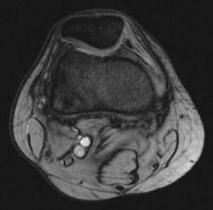
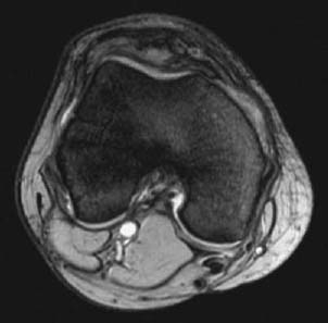
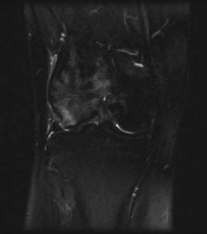
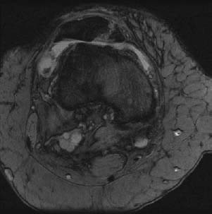
Radiologic Findings
Diagnosis
Related posts:
Stay updated, free articles. Join our Telegram channel

Full access? Get Clinical Tree


