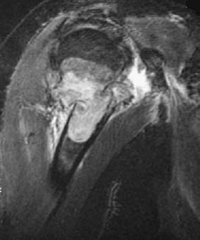
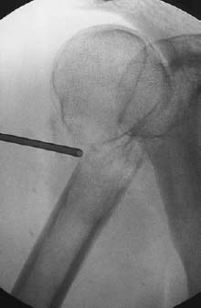
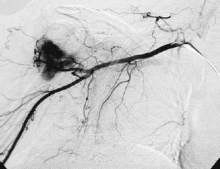
CASE 87 Hema N. Choudur, Anthony G. Ryan, and Peter L. Munk A woman with a known case of thyroid malignancy presented with acute right humeral pain with no history of trauma. She had tenderness and swelling along the proximal aspect of the arm with an inability to move her arm. Figure 87A Figure 87B Figure 87C Inversion recovery MRI (Fig. 87A) shows gross cortical dusruption through an oval high signal intensity lesion involving the neck and proximal shaft of the right humerus with secondary joint effusion. There is moderate medial displacement of the distal tibial fragment. Fluoroscopy (Fig. 87B) at the time of biopsy demonstrated a fracture of the surgical neck with a lucent lesion in the adjoining bone. The angiogram (Fig. 87C) revealed a dense hypervascular “blush” at the site of the fracture. Pathologic fracture, secondary to metastasis. Bone that is weakened by a preexisting condition, either primarily in the bone or secondary to a metastasis or primary tumor, can result in a fracture with trivial trauma. Usually the diagnosis is straightforward, given a history of malignancy or metabolic disease, but in other cases it may be the first indication of a malignancy. The histology and a directed search often lead to the primary malignancy. There are numerous conditions that weaken the bone, making it susceptible to fracture. These conditions can be classified as generalized disorders causing osteopenia and focal bony lesions.
Pathologic Fracture
Clinical Presentation


Radiologic Findings
Diagnosis
Differential Diagnosis
Discussion
Etiology
Pathophysiology
Clinical Findings
Stay updated, free articles. Join our Telegram channel

Full access? Get Clinical Tree


