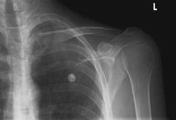
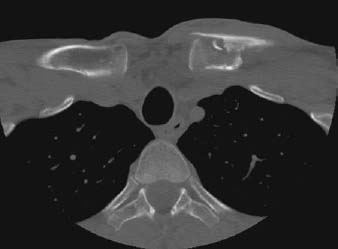
CASE 94 Hema N. Choudur, Anthony G. Ryan, and Peter L. Munk A young person presented to the emergency department with severe pain and swelling in the medial aspect of the left clavicle, after being assaulted. Figure 94A Figure 94B Figure 94C Plain radiographs of the chest (Fig. 94A) and left clavicle (Fig. 94B) show a subtle discontinuity of the interior cortex of the medial clavicle, suggestive of a fracture. The subsequent CT (Fig. 94C) confirms the minimally displaced fracture with no injury to the underlying structures. The sternoclavicular (SC) joint is seen to be congruent. Minimally displaced medial clavicular fracture. Traumatic fractures must be differentiated from pathologic and stress fractures, the latter especially in gymnasts. Isolated SC dislocations can be differentiated with the help of CT. Medial clavicular fractures are fairly uncommon, accounting for only 5% of clavicular fractures. It is important to diagnose them accurately in view of the important mediastinal structures posteriorly. The clavicle is an S-shaped bony strut that connects the shoulder girdle to the axial skeleton. It is the first bone to ossify in utero by intramembranous ossification, with the medial and lateral endochondral ossifications occurring later. The medial epiphysis appears after age 12 years; it is the last ossification center to fuse in the entire skeleton and occurs around age 23 to 25. Many SC dislocations in young athletes are instead injuries of the medial physis of the clavicle. Of interest is the fact that the SC joint capsule is thought to be the strongest in the body. The clavicle is attached securely to the medial aspect of the sternum and laterally to the scapula via the capsular and extra-articular ligaments, which maintain the width of the shoulders against gravity and muscle pull. The medial portion of the clavicle resists axial loading. It also protects the costoclavicular space, where the medial cord of the brachial plexus and the origin of the ulnar nerve are found. Fractures in this area put these structures at risk, as do clavicular nonunions and hypertrophic callus. The tubular shape of the medial clavicle and the flattened lateral clavicle place the medial clavicle at risk for fracture. In midclavicular fractures (Figs. 94D, 94E
Clavicular Fractures
Clinical Presentation

Radiologic Findings
Diagnosis
Differential Diagnosis
Discussion
Background
![]()
Stay updated, free articles. Join our Telegram channel

Full access? Get Clinical Tree


