CASE 97 Hema N. Choudur, Anthony G. Ryan, Peter L. Munk This patient presented to the emergency department with pain and swelling around the elbow with limitation of movement following a fall on an outstretched hand. Figure 97A Figure 97B Anteroposterior (AP) and lateral views of the elbow (Figs. 97A, 97B) were taken. The coranoid process of the ulna is disengaged from the trochlea, with the olecranon posteriorly dislocated. Note the small fracture of the coranoid process with soft-tissue swelling around the elbow joint. Posterior dislocation of the elbow. In adults, elbow dislocations are quite common, occurring second in frequency to shoulder dislocations. In children and adolescents, supracondylar fractures are more common. The inherently stable elbow joint requires a fair degree of force to cause dislocation; therefore, bony fractures accompany dislocations in a third of the patients. Posterior dislocations of the elbow are the predominant type and account for 80 to 90% of all elbow dislocations. However, anterior, lateral, or divergent dislocations can occur. A fall on an outstretched hand with the elbow in extension results in posterior dislocation of the elbow. Coexisting shoulder, wrist, and distal radioulnar joint dislocations may occur.
Elbow Dislocation
Clinical Presentation
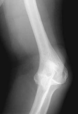
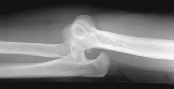
Radiologic Findings
Diagnosis
Differential Diagnosis
Discussion
Background
Etiology
Pathophysiology
Related posts:
Stay updated, free articles. Join our Telegram channel

Full access? Get Clinical Tree


