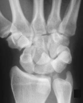
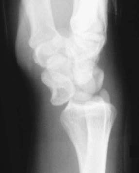
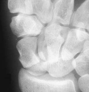
CASE 98 Peter L. Munk and Anthony G. Ryan A 36-year-old man was involved in a motorcycle accident. The vehicle spun out of control, and he struck the ground on an outstretched wrist. The patient complained of severe pain and inability to move the wrist freely. On examination, the wrist was swollen and had limited passive motion. Figure 98A Figure 98B Radiographs of the wrist show several abnormalities. On the anteroposterior (AP) view, a markedly abnormal appearance of the proximal carpal row is observed (Fig. 98A). The normal carpal arcs of Gilula are disrupted, and the lunate has assumed a triangular, or “slice-of-pie,” configuration. Examination of the lateral view reveals that the lunate has been displaced in the volar direction and is tilted anteriorly (Fig. 98B). No associated fractures are evident. Anterior dislocation of the lunate.
Lunate Dislocation
Clinical Presentation


Radiologic Findings
Diagnosis

Discussion
Background
Stay updated, free articles. Join our Telegram channel

Full access? Get Clinical Tree


