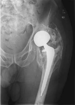
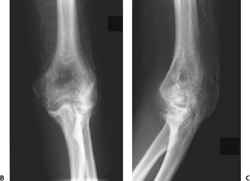
CASE 99 Hema N.Choudur, Anthony G.Ryan, and Peter L.Munk A 56-year-old man presented with pain and decreased mobility of the left hip, status posthemiarthroplasty. Figure 99A A conventional anteroposterior (AP) radiograph (Fig. 99A) of the left hip shows a left hemiarthroplasty with dense bony sheets and bars bridging the femoral shaft to the acetabular margins. Heterotopic ossification. None. Heterotopic ossification is abnormal true bone formation in the extraskeletal soft tissues. It has various synonyms, including myositis ossificans, a term no longer recommended, as it suggests a primary inflammation of the muscle (which has been shown histologically not to be the case) and as the condition also affects tissues other than muscle, such as fascia and tendons. Thirty percent of cases are said to occur in subcutaneous fat. It frequently occurs in traumatized soft tissues (after surgery, after spinal cord/brain injury, and following burns) but can also occur under other conditions causing muscle paralysis, such as poliomyelitis. Postsurgical heterotopic ossification is one of the most common causes of extraskeletal soft-tissue ossification and is seen following hip arthroplasty in 8 to 70% of cases. The process begins within 1 week of surgery. Cartilage is laid down by the second week, with trabecular bone formation evident by about the fifth week. Mature bone is formed after 6 weeks. It may regress and even disappear. Sometimes, the process may progress or halt. Initial pain and tenderness over the site of surgery may mimic cellulitis or a deep venous thrombosis (DVT). Later, an indurated hard mass becomes palpable. There are variable degrees of immobilization of the joint, depending on the amount of subsequent heterotopic bone formation. Trauma stimulates mesenchymal stem cells in the soft tissues, which then differentiate into osteoblasts, osteoid, and eventually heterotopic bone. Actual bone may be seen within the affected tissues. Following trauma, spindle cell proliferation may be seen during the first week. By 7 to 10 days, primitive osteoid develops at the periphery. Woven bone and primitive cartilage are laid down by about the second week. Trabecular bone forms by 2 to 5 weeks. Peripheral mature lamellar bone and central immature bone are seen after 6 weeks. Figures 99B, 99C AP (99B) and lateral (99C) radiographs of the right elbow show posttraumatic heterotopic bone formation along the fibers of the triceps muscle.
Heterotopic Ossification / Myositis Ossificans
Clinical Presentation
Radiologic Findings
Diagnosis
Differential Diagnosis
Discussion
Background
Clinical Findings
Pathology
GROSS
MICROSCOPIC
Imaging Findings
RADIOGRAPHY
Related posts:
Stay updated, free articles. Join our Telegram channel

Full access? Get Clinical Tree


