, Dennis M. Heisey1 and Glen E. Leverson1
(1)
Department of Surgery, School of Medicine and Public Health, University of Wisconsin, Madison, WI, USA
Abstract
This article describes a method for quantifying blood flow distribution among lung alveoli. Our method is based on analysis of trapping patterns of small diameter (4 μm) fluorescent latex particles infused into lung capillaries. Trapping patterns are imaged using confocal microscopy, and the images are analyzed statistically using SAS subroutines. The resulting plots provide a quantifiable way of assessing interalveolar perfusion distribution in a way that has not previously been possible. Methods for using this technique are described, and the SAS routines are included. This technique can be an important tool for learning how this critical vascular bed performs in health and disease.
Key words
LungBlood flowPerfusionInteralveolar pressureMicrospheres1 Introduction
Blood is arterialized in the lung because of diffusive exchange of oxygen and carbon dioxide between alveolar gas and capillary blood. This exchange operates effectively because the diffusion distance between the blood and the alveolar gas is only about 0.3 μm and because the combined surface area of the 3 × 10⁸ alveoli in the lung is about 70 m2 in humans or about one-fourth the size of a tennis court [1].
An important issue in efficient lung operation is the equal distribution of the blood flow to all of the alveoli. How the lung accomplishes this is unknown nor is it known if the capillaries control this perfusion distribution. This subject has received little attention because methods to study it have not previously existed.
To address this, we developed a method that is based on statistical analysis of the distribution of 4 μm diameter latex particles infused into the pulmonary circulation. These particles, which are only slightly larger than red cells, become trapped in alveolar capillaries, and their distribution is therefore an index of the blood flow distribution among the alveoli at the time of infusion. The following describes the details of this method, which we use in intact rats or isolated rat lungs to understand how blood flow is distributed among alveoli in health and disease [2–7].
2 Materials
1.
4 μm diameter rhodamine-labeled sulfate microspheres (FluoSpheres, Molecular Probes).
2.
Bovine serum albumin (Sigma).
3.
NaCl, Na2HPO4 (anhydrous), NaH2PO4∙2H2O.
4.
Laboratory rats fitted with acute or chronic indwelling femoral venous catheters.
5.
Infusion pump, rodent ventilator, and a source of compressed air to maintain lung inflation pressure at 5–10 cmH2O.
6.
Confocal fluorescence microscope for imaging rhodamine fluorescence.
7.
Personal computer equipped with SAS statistical software (SAS Institute, Cary NC) and NIH Image (National Institutes of Health) or Scion Image (Scion Corp.).
3 Methods
The following methods describe (1) preparation of the latex particles for infusion, (2) imaging the particles within excised, dried lungs, and (3) analysis of particle distributions within the lung.
3.1 Latex Particle Preparation
We infuse 4 μm diameter particles because more than 90 % of them become trapped within lung capillaries [2, 8]. Infusion of 3.6 μm particles results in only about 60 % retention. Thus, the larger particles are better markers of interalveolar perfusion distribution. It is worthwhile to infuse as many particles as possible to improve the resolution of the statistical analysis. However, if too many are infused they become crowded and cannot be imaged as separate particles, which degrades the analysis. We’ve found that 2 × 10⁸ is a good number to infuse into intact rats or isolated rat lungs. In vasoconstricted lungs, where the particles tend to cluster, 1 × 10⁷ is more appropriate.
We begin preparation of the infusion solution by adding the particle solution supplied by Molecular Probes to 5 ml of 0.5 % bovine albumin solution. For 2 × 10⁸ particles we add 204 μL; for 1 × 10⁷ we add 10 μL. We complete the solution by adding NaCl (0.043 g), Na2HPO4 (0.006 g), and NaH2PO4∙2H2O (0.002 g). The resulting solution consists of albumin-coated latex particles in phosphate buffered saline. The albumin is necessary to prevent the particles from clumping in the presence of the salts.
This solution is infused into the femoral venous catheter of an intact rat or into the pulmonary artery of isolated rat lungs over 1–2 min using an infusion pump. Once the infusion is complete, the lungs are removed and maintained at an inflation pressure of 20–25 cmH2O for at least 48 h, which causes the lungs to dehydrate. It is important that the lungs are never allowed to collapse after they are removed because the particles could redistribute.
3.2 Imaging the Particles
Air-dried lungs have the consistency of Styrofoam and are easily sliced using a razor blade. We cut four, 2–3 mm thick slices each from the left and right lungs. Three are parallel to the dorsal-ventral plane, and one is in the transverse plane parallel to the hilum [2].
Related posts:
 Confocal Imaging of Butterfly Tissue
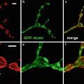 Confocal Imaging of Fluorescently Labeled Proteins in the Drosophila Larval Neuromuscular Junction
 Imaging Tools for Analysis of the Ureteric Tree in the Developing Mouse Kidney
 Live Confocal Analysis of Mutant- and Drug-Treated Drosophila Embryos
 Evaluating Confocal Microscopy System Performance
 Laser Scanning Confocal Microscopy: History, Applications, and Related Optical Sectioning Techniques
Stay updated, free articles. Join our Telegram channel

Full access? Get Clinical Tree


