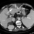
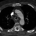
Fig. 5.1
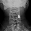
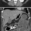
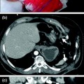
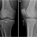
39-year-old woman with ACS as complication of 5° grade liver trauma. Bladder pressure is 35 mmHg measured by Kron method. a Scout CT image obtained 4 day after the surgery shows elevated right hemi diaphragm (white arrow) and packing (light blue arrow). b CT image shows big hematoma (white arrow) surrounding from “over packing” with hepatic segments lacerations with large ischemia (light blue arrow). c CT image shows retro hepatic cava vein is over-pressured and clotted (trombizzata) (light blue arrow). d CT image shows increased enhancement of the intestinal mucosa caused by venous congestion due to hypertension intra-abdominal (light blue arrow). e CT image shows increased anterior-posterior abdominal diameter studied at level where left renal vein crosses aorta (black arrow)
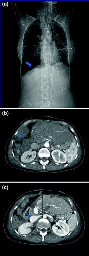
Fig. 5.2
The same patient after decompression surgery. a Scout CT image shows normal level of right hemi diaphragm and without packing (light blue arrow). b CT image shows thin fluid flap along shear section of the right liver (light blue arrow). c CT image shows the reperfusion of cava and renal veins (light blue arrow) and decreased of the anterior-posterior diameter
Follow-up CT scans showed the decompression effect of large open abdominal wound (Fig. 5.3).
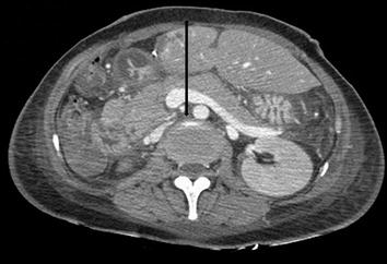

Fig. 5.3
Follow-up after 12 days. CT image shows decreasing of the anterior–posterior diameter with recovering of the ovoid shape
Stay updated, free articles. Join our Telegram channel

Full access? Get Clinical Tree


